Download
1 / 72
780 likes | 1.22k Views
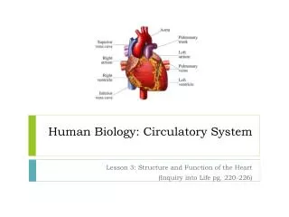
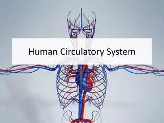
HUMAN CIRCULATORY SYSTEM. Circulatory Systems. Open system vs. closed system Both have a pump Open system has no blood vessels to hold blood Closed system has blood contained within vessels. Circulatory systems. The simplest of organisms do not have a circulatory system

E N D
Circulatory Systems • Open system vs. closed system • Both have a pump • Open system has no blood vessels to hold blood • Closed system has blood contained within vessels
Circulatory systems • The simplest of organisms do not have a circulatory system • Rely on diffusion/osmosis for regulation
Circulatory systems • Other simple organisms rely on the flow of water to exchange nutrients and wastes
Closed circulatory systems • Vary in complexity
Artery • Walls are thick • Muscular • Elastic, so they can stretch when heart contracts • Must withstand higher pressure than veins
Vein • Thin-walled (less muscle, less elastic tissue) • Contain VALVES to prevent back-flow
Capillaries • Blood vessels with the smallest diameter and thinnest walls
Capillary Beds • Exchange of carbon dioxide, oxygen, and nutrients occurs in the capillary beds
BLOOD PRESSURE • Measurement of both the pressure under which the heart contracts (systole), as well as the pressure under which the heart fills (diastole)
BLOOD PRESSURE • Blood pressure is checked in your arm, using a sphygmomanometer
Blood Pressure • The pressure generated by your heart can be measured in your arm because the pressure is transmitted through the muscular, elastic arteries
Blood Pressure • Measured in mm Hg • Reported as two numbers: systolic pressure diastolic pressure
Systolic blood pressure • Pressure transmitted when the left ventricle contracts • It is the pressure under which the blood is being forced out of the left ventricle into the aorta
Diastolic blood pressure • Is the filling pressure of the heart
Sounds of Korotkoff • Sounds of Korotkoff (are caused by turbulence in arterial blood flow. • A well-trained examiner can hear 5 different Korotkoff sounds, which vary slightly in quality • The first and fifth Korotkoff sounds are used to define blood pressure
Blood Pressure • Generally measured over the brachial artery
Why is blood pressure important? • Blood pressure must remain in a normal range so that tissues can receive adequate blood flow in order to exchange gases, nutrients, and wastes efficiently
HYPERTENSION • Elevation in blood pressure • Fairly common: about 30% • almost 1 in 3 adults! • HTN more common in • African Americans • Obese people
Hypertension • High blood pressure can damage the heart and blood vessels • Increases the chances of having a stroke • Cause is multi-factorial • Can be treated with medication; sometimes with diet and exercise alone
Electrocardiogram • Useful tool for studying the conduction of impulses through the heart
Electrocardiogram • P wave represents depolarization of the atrium • QRS complex represents depolarization of the ventricles • T wave represents repolarization (“recharging” for the next beat
ECG abnormalities • After a myocardial infarction (heart attack, “MI”), the damaged areas of the heart no longer have normal conduction of impulses • Results in a change in the normal waveforms
ECG abnormalities • Atrial fibrillation
ECG abnormalities • Acute myocardial infarction (heart attack)
COMPOSITION OF BLOOD • Blood is made up of a fluid portion and a cellular portion
PLASMA • Made up of water, proteins, ions, amino acids, sugars • Carries carbon dioxide (released from tissues) • Carries nutrients from the digestive system, hormones
Cells in the Blood • Erythrocytes: Red Blood Cells, RBC’s
Erythrocytes • Contain hemoglobin • A protein • Transports Oxygen
Erythrocytes • Hemoglobin structure
Erythrocytes • Live about 120 days • Produced in the bone marrow • Productions is regulated by a hormone from the kidneys- • erythropoetin
Blood Cells • Leukocytes = White Blood Cells • Function in the immune system • Form a second line of defense against bacteria and viruses • Larger than RBC’s
Leukocytes • Granulocytes • Neutrophils • Basophils • Eosinophils • Lymphocytes • Macrophages