Download
1 / 81
830 likes | 1.61k Views
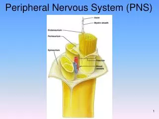
Peripheral Nervous System (PNS). Spinal Nerves Posterior View. Cut nerves. If a small nerve is cut, it will regenerate because where are the cell bodies? In the posterior root ganglion (sensory) or anterior horn (motor). Since the cell body is about a meter away, axons can regrow.

E N D
Cut nerves • If a small nerve is cut, it will regenerate because where are the cell bodies? In the posterior root ganglion (sensory) or anterior horn (motor). • Since the cell body is about a meter away, axons can regrow. • Large nerves are harder to regrow, but you can still stitch the ends together at the epineurium and perineurium, and you may get healing.
Doctors trying to fix damaged nerves • http://www.foxnews.com/health/2012/11/13/doctors-trying-to-fix-damaged-nerves/ • Nerve disease and injuries are tough to treat, largely because there's no way to regenerate many damaged nerve cells. Neurologist Joseph Corey is trying to change that. • Corey and a team of scientists used tiny polymer fibers as a scaffold. They coaxed an oligodendrocyte to form a myelin sheath around the fiber. The artificial fiber mimicked an axon. • Myelin provides the pathways along which some nerve cells regenerate after an injury. When a person has MS or cerebral palsy, the oligodendrocytes are damaged and don't function properly and the myelin sheaths start to break down.
Rat Brain in a Dish Flies Plane • An electrode grid was placed at the bottom of a glass dish and then covered with rat neurons that gradually formed a neural network -- a brain. • They then used the brain to control an F-22 fighter jet flight simulator. • The research could lead to tiny, brain-controlled prosthetic devices and unmanned airplanes flown by living computers.
Exoskeleton Helps Paralyzed Patients Walk • Exoskeletons have been designed for military use and boosting strength. But the same technology that makes people able to lift heavier loads might also one day allow those with spinal injuries to walk. • Ekso Bionics, a California company, developed the Human Universal Load Carrier, or HULC for the military, and another one called the Ekso, for people who need either physical therapy or rehabilitation.
Pinched nerves • When a nerve gets pinched (e.g. herniated disc), it damages the nerve by interfering with its action potential, causing weakness, pain, or paralysis.
Tens unit • Transcutaneous electrical nerve stimulation TENS is usually applied at high frequency with an intensity below motor contraction. It just blocks pain impulses. This is a different machine than a muscle stimulator, but they look the same. TENS is only available by Rx, but muscle stimulators can be sold without a Rx.
Disruption of Blood Supply • When a body part “falls asleep”, the region has become ischemic (lack of blood flow), impairing the action potential of the nerves. Unlike the CNS, when blood is restored to the PNS, the nerves recover. Damage to the CNS tends to be permanent, but damage to the PNS tends to heal.
DAMAGE TO THE NERVOUS SYSTEM • If a person has a spinal cord injury in their cervical region, they could have quadriplegia (arms and legs paralyzed). • If a person has a spinal cord injury in their thoracic region, they could have paraplegia (just legs are paralyzed).
SOME CLINICALLY IMPORTANT PERIPHERAL NERVES: • Note: an epidural nerve block during child birth will numb the mother from her navel to her knees. • PUDENDAL NERVE: this is the nerve that can be anesthetized during childbirth as an alternative to an epidural (a pudendal nerve block is also called a saddle block because the numb areas are where you would be touching a saddle). • PHRENIC NERVE: allows the diaphragm to contract. If it gets severed, the person can no longer breathe without assistance.
SENSORY CUTANEOUS NERVES • These come out of the spinal cord and go to specific regions of skin on the body. • For example, nerve C4 innervates the skin region C4 of the DERMATOME MAP. • It’s important to know these dermatome map regions (not for this class), especially physical therapists and nurses.
Dermatome Map • If a patient has a shooting pain down the anterior shin, what nerve is pinched? L5. • Numbness in pinky and ring finger is what nerve? C8. • If a workman’s comp patient comes in saying his whole hand is numb, no other symptoms, you know he’s lying because the nerves don’t run that way.
Nerve Plexus A PLEXUS is a network of nerves that primarily serves the limbs. There are four major plexi: cervical, brachial, lumbar, and sacral. 1. CERVICAL PLEXUS comes out of the neck and are cutaneous nerves (sensory input of the skin) of the neck and back of the head. The phrenic nerve (supplies the diaphragm) is also in this plexus.
BRACHIAL PLEXUS 2. BRACHIAL PLEXUS • This is the major group of nerves that supply the upper limbs. It runs through the axilla. • If a person leans their armpits on their crutches, they can damage this plexus and lose the use of their arms. • The nerves in the brachial plexus change names as they go to different regions in the arm.
Major Nerves of the Upper Extremity Axillary Musculocutaneus
Axillary Nerve • Deltoid
Musculocutaneus Nerve • Supplies anterior muscles of the arm
Median Nerve • Supplies no muscles of the arm • Supplies anterior forearm (except flexor carpi ulnaris) • Damage can cause • Hand of benediction • Ape Hand • Carpal Tunnel Syndrome Patient trying to make a fist
MEDIAN NERVE: Ape Hand • This is the nerve that gets cut when people try to slit their wrists. • The arteries are so small in the wrist; people rarely die from this type of suicide attempt. • However, they live with a lot of tissue damage. • They are not able to move the thumb towards the little finger, so it is hard to pick up small objects. • This is called “ape hand”.
Median Nerve:Carpel Tunnel Syndrome • The median nerve travels under the transverse carpal ligament. • The nerve is pinched in carpal tunnel syndrome.
Patient Case • George has been a computer programmer for 20 years. He has numbness in his right hand on the thumb, index finger, and middle finger. • Tapping on the carpal tunnel causes parathesias (tingling) in the median nerve distribution (positive Tinel’s sign). • Placing his wrist in sustain flexion for one minute also causes the parathesias (positive Phalen’s test).
Patient Case • Treatment began with splinting the wrist in neutral position and patient education for proper ergonomics (use a wrist pad while typing).
Trigger Finger • Trigger finger is one example of the disability that can be created when repetitive trauma to a flexor tendon results in the formation of nodules on the tendon. Finger flexion may be prevented completely, or the finger may be unable to re-extend.
Ulnar Nerve • Supplies flexor carpi ulnaris • “Funny Bone” • Damage can cause claw hand; cannot adduct or abduct fingers
Radial Nerve • Supplies muscles on the posterior arm and forearm • Triceps brachii • Extensor carpi radialis • Extensor digitorum communis • Damage can cause wrist drop • Also called “waiter’s hand”
Carpel Tunnel Syndrome Ape Hand
Axillary, Musculocutaneus, Ulnar, Median, Radial, Nerves Figure 14.4
Damage to Brachial Plexus Congenital (brachial plexus damaged during birth) Klumpke’s paralysis Acquired Brachial Plexus injuries Crutch paralysis (total upper extremity paralysis) Claw Hand Carpal Tunnel Syndrome, Ape hand, Hand of benediction Wrist Drop (Waiter’s Hand) Brachial Plexus
LUMBAR PLEXUS 3. LUMBAR PLEXUS • FEMORAL NERVE is the main nerve to the anterior thigh.
Lumbo-Sacral Plexus • Lumbar: • Femoral nerve • Sacral: • Sciatic nerve
The Lumbar Plexus Figure 14.15
Sacral Plexus 4. SACRAL PLEXUS are spinal nerves from L4-S5 • Some of the fibers from the lumbar plexus mix with the sacral plexus, so these are often referred to together as the lumbosacral plexus. • SCIATIC NERVE is the largest branch of the sacral plexus and the largest nerve in the body; it leaves the pelvis through the greater sciatic notch. • A short, thick muscle (Piriformis muscle) covers the greater sciatic notch, and when it contracts, it can pinch the sciatic nerve, causing a type of sciatica (sciatic nerve irritation) known as piriformis syndrome. • This can be alleviated by stretching exercises. However, sciatica can also be caused if there is a herniated lumbar disc, in which case stretching exercises make it worse.
The Sacral Plexus Figure 14.16a, c
Spinal steroid shots may have little effect on sciatica • http://www.foxnews.com/health/2012/11/13/spinal-steroid-shots-may-have-little-effect-on-sciatica/ • For the back pain component of sciatica, the researchers found that the injections didn't seem to make a difference over short or long periods of time. • When it came to leg pain, there was no difference a year or so after the injection, but there was a statistically significant drop in pain scores over the short term - about 2 weeks to 3 months.
Nerves of the Lower Extremity Obturator Femoral The sciatic nerve supplies the back of the thigh, then branches out into the TIBIAL and FIBULAR (peroneal) nerves, which supply the leg and foot. The fibular nerve branches into superficial and deep.
Lower Extremity Nerves • Obturator Nerve • Supplies adductor muscles • Femoral Nerve • Supplies anterior Thigh • Sciatic Nerve • Supplies back of thigh • Tibial Nerve • Supplies posterior leg and foot • Common Fibular Nerve • Superficial branch • Supplies lateral side of leg • Deep branch • Supplies anterior leg • Injury causes “Foot Drop”
Tibial Nerve • Sometimes a small branch of the tibial nerve in the foot gets pinched between the metatarsal heads, and the irritation causes nerve swelling and pain. • It is called a neuroma (“nerve tumor”) and manifests as pain in the ball of the foot, made worse with high heels.
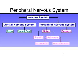
Nervous System Classification • Somatic Nervous System • Sensory nerves (somatosensory neurons) • Reflexes • Sensory, interneurons, lower motor neurons • Motor nerves to skeletal muscle (somatic motor neurons) • Upper and lower motor neurons • Autonomic Nervous System • Motor nerves to smooth and cardiac muscle (visceral motor neurons) • Sympathetic • Parasympathetic
AUTONOMIC NERVOUS SYSTEM We don’t have voluntary control over these nerves. They are involved digestion, blood flow, urination, defecation, glandular secretion. Therefore, the ANS supplies the glands, smooth muscle, and cardiac muscle, but NOT the skeletal muscle. For this reason, the ANS is also called the general visceral motor system.