Download
1 / 1
10 likes | 119 Views
Histopathological evaluation of adipose-derived mesenchymal stem cells on cartilage defects of knee in male rabbits. M.Babazadeh 1 , N.Tanideh 2 , D.Mehrabani 2 , M.J. Ashraf 3 , S.Hoseinzadeh 4 , A.tamaddon 1 , M.Heidari 1 , O.Kuhi 5 , F.Parvin 2 .

E N D
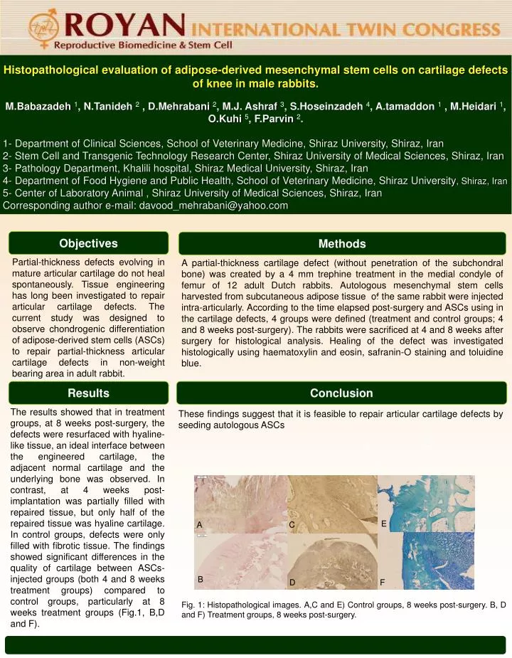
Histopathological evaluation of adipose-derived mesenchymal stem cells on cartilage defects of knee in male rabbits. M.Babazadeh1, N.Tanideh2 , D.Mehrabani2, M.J. Ashraf 3, S.Hoseinzadeh4, A.tamaddon1 , M.Heidari1, O.Kuhi5, F.Parvin2. 1- Department of Clinical Sciences, School of Veterinary Medicine, Shiraz University, Shiraz, Iran 2- Stem Cell and Transgenic Technology Research Center, Shiraz University of Medical Sciences, Shiraz, Iran 3- Pathology Department, Khalili hospital, Shiraz Medical University, Shiraz, Iran 4- Department of Food Hygiene and Public Health, School of Veterinary Medicine, Shiraz University, Shiraz, Iran 5- Center of Laboratory Animal , Shiraz University of Medical Sciences, Shiraz, Iran Corresponding author e-mail: davood_mehrabani@yahoo.com Objectives Methods Partial-thickness defects evolving in mature articular cartilage do not heal spontaneously. Tissue engineering has long been investigated to repair articular cartilage defects. The current study was designed to observe chondrogenic differentiation of adipose-derived stem cells (ASCs) to repair partial-thickness articular cartilage defects in non-weight bearing area in adult rabbit. A partial-thickness cartilage defect (without penetration of the subchondral bone) was created by a 4 mm trephine treatment in the medial condyle of femur of 12 adult Dutch rabbits. Autologous mesenchymal stem cells harvested from subcutaneous adipose tissue of the same rabbit were injected intra-articularly. According to the time elapsed post-surgery and ASCs using in the cartilage defects, 4 groups were defined (treatment and control groups; 4 and 8 weeks post-surgery). The rabbits were sacrificed at 4 and 8 weeks after surgery for histological analysis. Healing of the defect was investigated histologically using haematoxylin and eosin, safranin-O staining and toluidine blue. A Results Conclusion The results showed that in treatment groups, at 8 weeks post-surgery, the defects were resurfaced with hyaline-like tissue, an ideal interface between the engineered cartilage, the adjacent normal cartilage and the underlying bone was observed. In contrast, at 4 weeks post-implantation was partially filled with repaired tissue, but only half of the repaired tissue was hyaline cartilage. In control groups, defects were only filled with fibrotic tissue. The findings showed significant differences in the quality of cartilage between ASCs-injected groups (both 4 and 8 weeks treatment groups) compared to control groups, particularly at 8 weeks treatment groups (Fig.1, B,D and F). These findings suggest that it is feasible to repair articular cartilage defects by seeding autologous ASCs A B C E A C B D C D B D F Fig. 1: Histopathological images. A,C and E) Control groups, 8 weeks post-surgery. B, D and F) Treatment groups, 8 weeks post-surgery.