Download
1 / 1
10 likes | 141 Views
Phase 1. Flashing Star. 1000 mHz tone. Pt opens box and pushes button with left hand to start trial. Visual/Auditory Cue. Intentional Act. Phase 2. If incorrect response, pt repeats word while gesturing with left hand. Pt names black-and- white drawing centered on monitor.

E N D
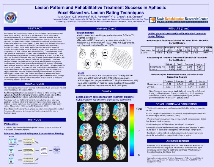
Phase 1 Flashing Star 1000 mHz tone Pt opens box and pushes button with left hand to start trial Visual/Auditory Cue Intentional Act Phase 2 If incorrect response, pt repeats word while gesturing with left hand Pt names black-and- white drawing centered on monitor Flashing Star Pt opens box and pushes button with left hand to start trial Visual Cue Intentional Act Phase 3 Flashing Star Pt initiates trial with nonsymbolic circular left hand movement Visual Cue Generalizable Intentional Act Lesion Pattern and Rehabilitative Treatment Success in Aphasia: Voxel-Based vs. Lesion Rating Techniques M.A. Cato1, C.E. Wierenga2, R. B. Parkinson3, Y.-L. Chang3, & B. Crosson3 1Nemours Children’s Clinic, Jacksonville, FL; 2VA San Diego Healthcare System and University of California, San Diego 3 University of Florida and the Malcom RandallVA RR&D Brain Rehabilitation Research Center; Gainesville, FL ABSTRACT Methods (Cont.) RESULTS (Cont.) • Lesion Ratings • Lesion extent was rated in grey and white matter ROI’s on T1-weighted MRI scans. • The selected ROI’s and rating schema were adapted from Naeser et al.’s methods (1989; 1990; 1998), with supplemental use of an additional atlas (Matsui, 1978). Lesion pattern corresponds with treatment outcome-Lesion Ratings Factors that predict recovery potential in chronic nonfluent aphasia are not well understood. Recently, Crosson et al. (Richards et al., 2002) developed a rehabilitative treatment of language production deficits in aphasia. Here, we examine the relationship between lesion pattern and level of success on this treatment using two methodologies, qualitative lesion ratings and quantitative voxel-based lesion-symptom mapping (VLSM). Previously, we found that level of pre-treatment comprehension positively correlated with level of treatment success (Cato et al., 2004). Here, we hypothesized that level of treatment success is related to lesion extent in posterior regions, given their importance in comprehension. Both analyses were performed using T1-weighted MRI scans. The qualitative analysis was modeled after Naeser et al.’s methods (e.g., 1998). VLSM analyses were performed according to Bates et al. (2003). Nine left MCA nonfluent aphasia patients with wide ranging lesion patterns were included in this analysis. Results from both methods confirmed our hypothesis. Qualitative analysis revealed that treatment change scores were significantly negatively correlated with lesion extent in several posterior regions, including Wernicke’s area, posterior limb of the internal capsule, and the posterior 1/3 of periventricular white matter (PVWM). Similar results were found using VLSM. However, results of VLSM revealed some additional anterior regions that were associated with negative treatment outcome. These anterior regions included the left inferior frontal gyrus, insular cortex, and anterior periventricular white matter areas. Thus, while the 2 lesion analysis methodologies provided similar results, findings were not wholly convergent. Further comparison of qualitative vs. quantitative lesion rating methods is warranted. Relationship of Treatment Outcome to Lesion Size in Posterior Cortical Regions Relationship of Treatment Outcome to Lesion Size in Anterior Cortical Regions • VLSM • A mask of the lesion was created from the T1-weighted MRI scans using ROI tools within the AFNI software package. • VLSM algorithms programmed in MATLAB (The Mathworks, MA) were used to create a T-map for voxels significantly associated with poor treatment response across the 9 participants. Relationship of Treatment Outcome to Lesion Size in Subcortical Regions BACKGROUND The factors that predict recovery potential in chronic nonfluent aphasia are not well understood (Basso & Farabola, 1997). Past findings indicate worse recovery is related to initial severity of the aphasia (particularly initial comprehension ability), lesion size, and the presence of posterior lesions, particularly in Wernicke’s area. Previously we found that level of comprehension, but not lesion size, was positively correlated with level of treatment improvement. Here, we present findings on lesion pattern and treatment response using two methods, lesion ratings (Naeser et al., 1998) and VLSM (Bates et al., 2003). Hypothesis: In chronic nonfluent aphasic patients, both methods will reveal that lesion extent in posterior brain regions will be inversely related to level of treatment gains. Results Note: Treatment improvement (rpch_ip3) defined as (Mean%Correct Probeph3 – Mean%Correct Probebl)/(100-Probebl).). PLIC = posterior limb internal capsule; PSM = posterior supramarginal;PVWM = periventricular white matter; GP = globus pallidus. Lesion pattern corresponds with treatment outcome-VLSM Posterior regions most significantly associated. CONCLUSIONS and DISCUSSION • Treatment response was most adversely affected by lesions in posterior regions. • In this sample comprehension performance was positively correlated with treatment improvement (Cato et al., 2004). • Posterior lesion involvement may correspond with comprehension deficits that dampen treatment gains. • Lesion rating and VLSM methods were largely convergent. • Drawback of voxel-based method: requires accurate appraisal of lesion vs. no lesion in each voxel; also optimal with very large sample sizes. • Drawback of rating methods include requirement of expert raters, time demands, and subjective nature of rating level of lesion. 3.06 METHODS Participants 9 chronic, right-handed nonfluent aphasic patients (4 male, 5 female; 8 Caucasian, 1 African American) Intention Treatment to Improve Confrontation Naming t Acknowledgments We would like to acknowledge Zachary Cook and Sheila Rosenthal for their assistance with this project. Infrastructure and technical support generously provided by the UCSD and San Diego VA Laboratory of Cognitive Imaging, San Diego ,CA. Address for correspondence: M. Allison Cato Jackson, Ph.D., Nemours Children’s Clinic, Neurology Division, 807 Children’s Way, Jacksonville, FL 32207. E-mail: acato@nemours.org 0