Download
1 / 68
690 likes | 1.57k Views
Gingival and Dentogingival Junctional Tissues DHY114 – Week 2. Chapter 10. Gingival Tissues. Gingiva Attached Free Interdental gingiva Col Gingival margin Free gingival groove. Gingiva. Free and attached gingiva. Attached Gingiva. Brown Spotted with brown (Melanin pigmentation).

E N D
Gingival and Dentogingival Junctional TissuesDHY114 – Week 2 Chapter 10
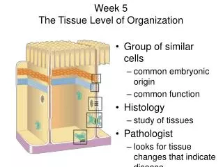
Gingival Tissues • Gingiva • Attached • Free • Interdental gingiva • Col • Gingival margin • Free gingival groove
Gingiva • Free and attached gingiva
Attached Gingiva • Brown • Spotted with brown (Melanin pigmentation)
Free Gingiva • Covered withkeratinizedepithelium • Interdental papilla • Free gingival groove • Sulcus
Attached Gingiva • Keratinizedepithelium • Pink color • Variations of gingiva found in dark skinned individuals (Asians, African Americans, Indians, and people of the Mediteranean (Italians, Arabs, Turks, Jewish, and Yesmenites) • Brown • Spotted with brown (Melanin pigmentation) • Sulcus
Anatomy of Periodontium Darby. Mosby’s Comprehensive Review of Dental Hygiene, Mosby, 5th ed. 2002 p. 525
Nonkeratinization • Col: (kawl) Depression between the lingual and facial papilla under the contact area. The probe readings Underneath the col may give the first sign of periodontal disease. Wilkins. Clinical Practice of the Dental Hygienist, 8th Ed., 1999, Lippencott, Williams and Wilkins, p. 192.
Periodontium: Cementum, Alveolar Bone, Periodontal LigamentDHY114 – Week 2 Chapter 14
Periodontium • Cementum • Alveolar bone • Periodontal ligament • Gingiva • Purpose: support of teeth
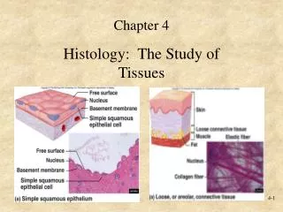
Cementum • Not always visible • Hard tissue • Thickness varies • Cementoenamel junction
Cementum • Functions to seal tubules of the root and provides attachment for the principal fiber groups. • Thinner at the cervical area than the apical portion of the root. • Thin layer of calcified connective tissue
Sharpey’s Fibers • At the very ends of the periodontal ligament (PDL), which are collagen fibers Smith & Harst. Head and Neck Histology & Anatomy, Appleton and Lange, 2000 p. 192.
Cementum • The cementum does NOT meet enamel at the CEJ 10% of the time. Wilkins. Clinical Practice of the Dental Hygienist, 8th Ed., 1999, Lippencott, Williams and Wilkins, p. 229.
Cementum • Acellular : Cervical 1/3 • Cellular: Apical 2/3, adds new layers • Functions to seal tubules of the root and provides attachment for the principal fiber groups.
Cementum • Cellular cementum is most likely found: • At the apical portion of the tooth • At the CEJ • Equally throughout the root surfaces • Closest to the dentin
Cementum • Cellular cementum is most likely found: • At the apical portion of the tooth • At the CEJ • Equally throughout the root surfaces • Closest to the dentin
Cementum • At which location does cementum have the greatest thickness? • At mesial root surfaces • At Sharpey’s attachment sites • Closest to the CEJ • At the apex or furcation areas
Cementum • At which location does cementum have the greatest thickness? • At mesial root surfaces • At Sharpey’s attachment sites • Closest to the CEJ • At the apex or furcation areas
Portion of the maxilla Alveolar bone Basal bone
Periapical Occlusal
Trabecular Bone, Alveolar Crest, and Lamina Dura Smith & Harst. Head and Neck Histology & Anatomy, Appleton and Lange, 2000 p. 199.
Alveolus, Alveolar bone proper, Cribriform plate, Lamina dura (radiographically) Brand and Isselhard. Anatomy of Orofacial Structures. 7th Ed. 2003, p. 15, Fig. 2-6.
Lamina Dura Langland, Langlais, and Preece. Principles of Dental Imaging, 2nd ed. Lippincott Williams, And Wilkins, 2002 p. 412.
Alveolar Process Woelfel and Scheid. Dental Anatomy 6th Ed. 2002, Lippincott, Williams and Wilkins. p. 15.
Periodontal Ligament • Acts as a shock absorber
Sharpey’s Fibers • At the very ends of the periodontal ligament (PDL), which are collagen fibers Smith & Harst. Head and Neck Histology & Anatomy, Appleton and Lange, 2000 p. 192.
Principal Fibers Gingival fiber Nelson. Saunders Review of Dental Hygiene. Saunders, 2000, p. 5
Principal Fibers Gingival fiber Darby. Mosby’s Comprehensive Review of Dental Hygiene, Mosby, 5th ed. 2002 p. 527.
Principal Fibers • Identify the following: A B E C D Darby. Mosby’s Comprehensive Review of Dental Hygiene, Mosby, 5th ed. 2002 p. 527.
Principal Fibers • Identify the following: A- Alveolar crest B - Horizontal E - Interradicular C - Oblique D - Apical Darby. Mosby’s Comprehensive Review of Dental Hygiene, Mosby, 5th ed. 2002 p. 527.
Mixture of Principal and Gingival Fibers • List the following fibers in A and B A, B, and C= Gingival AC H O AP TS Gingival fiber Brand and Isselhard. Anatomy of Orofacial Structures. 7thth Ed. 2003, p.81.
Mixture of Principal and Gingival Fibers • List the following fibers in A and B A, B, and C= Gingival AC- Alveolar crest H- Horizontal O- Oblique AP-Apical TS- Transseptal Gingival fiber Brand and Isselhard. Anatomy of Orofacial Structures. 7thth Ed. 2003, p.81.
Principal Fibers All Have Obtained Adequate Information for you to do well with the Principal fiber groups AC- Alveolar crest H- Horizontal O- Oblique AP-Apical I - Interradicular Darby. Mosby’s Comprehensive Review of Dental Hygiene, Mosby, 4th ed. 1998 p. 452.
Principal Fibers and a Gingival Fiber Gingival fiber Nelson. Saunders Review of Dental Hygiene. Saunders, 2000, p. 5
Gingival Fibers: Transseptal • From the cervix of one tooth to the cervix of the adjacent tooth, along crestal bone Smith & Harst. Head and Neck Histology & Anatomy, Appleton and Lange, 2000 p. 208.
Rete Pegs • Folds that appear in the gingiva (lamina propria) that look like fingers or ridges. They are made up of epithelial tissues to strengthen the tissue. Smith & Harst. Head and Neck Histology & Anatomy, Appleton and Lange, 2000 p. 208.
Gingival Fibers Transseptal fibers on next slide Darby. Mosby’s Comprehensive Review of Dental Hygiene, Mosby, 5th ed. 2002 p. 526.