Download
1 / 11
270 likes | 928 Views
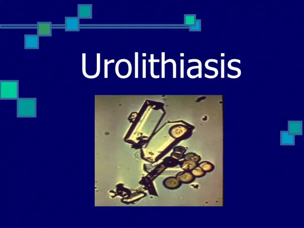
Urolithiasis. Presentation by melissa vandyke. What is urolithiasis?????. a formation of urinary calculi in any area of the urinary tract.

E N D
Urolithiasis Presentation by melissa vandyke
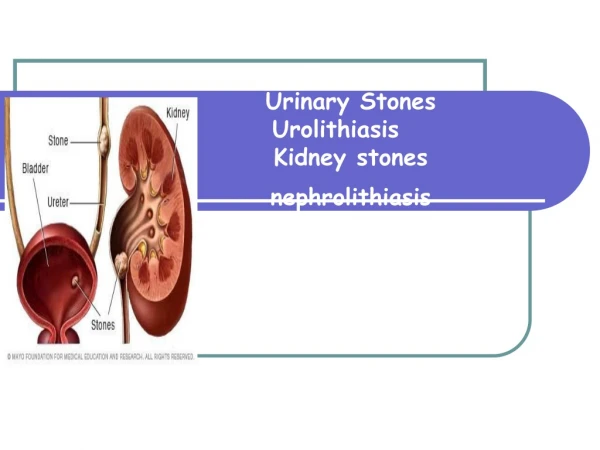
What is urolithiasis????? • a formation of urinary calculi in any area of the urinary tract. • Named specifically to indicate where they are located or formed: nephrolithiasis (stones in the kidney), ureterolithiasis (stones in the ureter), and cystolithiasis (stones in the bladder) • Urolithiasis develops from minerals that have precipitated out of solution and adhere, forming stones that varie in size and shape • Some people are predisposed; people that are immobile, are hyperparathyroid, people that have recurrent UTI’s, • Also individual history and some foods, nutrients, and medications also contribute to development of stones.
Clinical Manifestations…. • Size and degree of mobility of the stone influences what symptoms you might have • Patients with renal colic will receive attention immediately whereas a patient with a less mobile stone with wait until there are signs and symptoms of infection to seek assistance
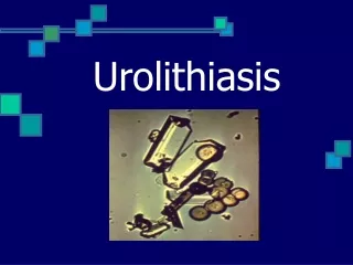
A look at stones……….. OUCH!!!!
Assesment of a patient with urolithiasis…….. • Patient with mobile calculi will complain of intractable pain and is usually accompanied by nausea and vomiting • Patients has pain that starting in the flank and radiating into the groin, genitalia, and the inner thigh. • Patient with a less mobile stone will have signs and symptoms as that with a UTI ( pain and burning with urination, nocturia, abdominal discomfort, flank pain, hematuria, or pyuria.) • Collection of objective data would include assessing for presence of hematuria and vomiting
Diagnostic Test………. • KUB and IVP/IVU radiography, ultrasound, cystoscopy, and urinalysis. • Other test may also be ran to determine stone content, presence of infection, and alterations in blood chemistry that may influence stone formation. • 24 hour urine examination may be done to detect abnormal excretion of calcium oxalate, phosphorus, or uric acid.
Medical Management…… • Antiinfective agents may be administerd in the presence of infection • Stones may need to be removed surgicaly (ureterolithotomy, pyelolithotomy, nephrolitomy) • Chemolytic agents (alkylating or acidifying agents) may be instilled to dissolve stones. • Lithotrispy- patient is submerged in a special tank of water and ultrasonic shock waves are used to pulverize the stone. (urine is still strained) • Long term management may include dietary adjustments to influence the urine pH or to decrease availabilty of certain substances to discourage stone formation • Moderate reduction of calcium phosphorus and purine-containing foods when stones are caused by metabolic abnormalites.
Medical Management con’t…. • Adequate daily fluid intake of 2000ml will help cleanse urinary tract • Avoid foods such as cheese, greens, whole grains, carbonated beverages, nuts, chocolate, shellfish, and organ meat • In calcium stone formation, sodium cellulose phosphate binds with ingested calcium and prevents its absorbtion • Aluminum hydroxide gel with bind with excess phosphorus allowing intestinal excretion rather than urinary excretion • Allopurinol (Zyloprim) reduces serum nitrate levels
Nursing Interventions……. • The patient should remain active and increase fluid intake (at least 2000mL per day) • STRAIN ALL URINE!!!!! • Asses urine for hematuria • Monitor BUN and creatinine for indications of continuing urinary obstruction • People that are calcium stone formers should avoid dairy products and antiacids • Avoid foods such as cheese, greens, carbonated beverages, whole grains, organ meats, chocolate, and nuts.