Download
1 / 30
310 likes | 375 Views
Explore the intricate mechanisms influencing blood flow regulation, including metabolic product effects, vessel diameter changes, and the roles of myogenic and neuro-humoral pathways. Understand the basal tone of vessels, smooth muscle actions, and central nervous system involvement in maintaining vascular function.

E N D
Influencesof metabolic products on the diameter of vessels Increase of Н+ ions, pyroveniger and lactic acids, decrease of pO2 and increase ofpCO2in tissues Decrease of tone of precapillary sphincters Dilatation of arterioles Increaseof blood flowin organs
Basal tone of vessels Smooth muscles of vessels wall don’t relax whole. It all time has some tension – muscular tone. Tonic condition is connect with changes of electrical characteristic and some contraction of muscles. Tone of smooth muscles support by two mechanisms: myogenic and neuro-humoral. Miogenic regulation play the main role in the support of vessel tone. When absent all nervous and humoral influences, present vessel tone or basal tone. In the base of basal tone is possibility of some smooth cells to the spontaneously activity and spread of excitation from cell to cell;it provide rhythmical changing of tone. It present in arterioles, precapillares sphincters. Influences, which decrease level of membrane potential, increase frequency of spontaneously impulses and amplitude of contraction of smooth muscles. Hyper polarization of membrane leads to disappeared of spontaneously excitability and muscles contraction.
Role of mechano- and chemoreceptorin regulation of the vessels tone From mechanoreceptors of aorta arc sensory information transmit by left depressor (aortic)nerve, brunch of n.vagus to the medulla oblongata. Excitation from mechanoreceptorsof carotid sinus zone lead by Sino carotid nerve(brunchof glossopharingeal nerve) to the medulla oblongata.
Characteristic of afferent link Sensory innervations of heart and vessels is present by nerve ending. Receptors divided by it function on mechanoreceptors, which are reacted on the changing of arterial pressure and chemo receptors, which are reacted on the changing ofchemical composition of blood. Irritation for mechanoreceptors is the speed and level of tissues stretching by increase or pulse wave of blood pressure. Angioreceptors are present at all vessel system and have the whole receptor field, it maximal presents at the main reflector zones: aortic, sino-carotid, in the vessels of pulmonary cycle of the blood circulation. At the answer on the each systolic increase of arterial pressure, mechanoreceptors of that zones generate impulses, which disappeared in the diastolic decrease of pressure. Minimal threshold of excitation of mechanoreceptors is 40 mm Hg, maximalis 200 mm Hg. Increase of pressure higher than that level don’t lead to addition increase of impulsation.
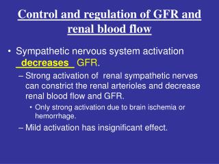
Central part in regulation of vascular tone Central mechanisms, which regulate connection between level of cardiac output and tone of vessels, working by help of complex of nervous structures, which named vasomotor center. Structures of vasomotor center are present in spinal cord, medulla oblongata, hypothalamus, cortex of big hemisperes. Spinal level of regulation is in the lateral root of thoracic and lumbar segments and consist of nervous cells, axons of which produce the vasculoconstrictors fibers. That neurons support their level of excitation by help of impulses from higher structures of nervous system.
Vasomotor center of medulla oblongatais the main center of regulation of blood flow.It located on the bottom of 4 ventricle, in it upper part.Vasomotor center divided on pressor and depressor zones. Pressor zone support increase of arterial pressure. It connect with the increase of tone of resistive vessels. Also increase frequency and strength of heart contractionand as result minute volume of blood flow. Regulatory influences of neurons of pressor zoneact by help ofincrease of tone of sympathetic nervous system on heart and vessels. Depressor zone support decrease of arterial pressure, heart work. It is the place of changes the impulses, which are coming from mechanoreceptors of reflector zones and cause central inhibition of tonic impulses of vasoconstrictors. Parallel the information from that zone by help of parasympathetic nerves go to heart. As result, decrease work and stroke volume of blood. Also, depressor zone act reflector inhibition of pressor zone.
Role of brain cortexand hypothalamus in regulation of blood flow Centers of hypothalamus give the descendent influences on the vasomotor center of medulla oblongata. In hypothalamus present depressor and pressor zones. That is why hypothalamiclevelgive the same double reaction as bulbar center. Posterolateral part of hypothalamus cause excitation of vasomotor center. Anterior part of hypothalamus can cause mild inhibition of one. Some zones of cortexalso give the descendent influenceson the vasomotor center of medulla oblongata.Motor cortex excites vasomotor center. Anterior temporal lobe, orbital areas of frontal cortex, cingulated gyrus, amygdale, septum and hippocampus can also control vasomotor center. That influencesformas a resultof comparethe information, whichenter in higherpartof nervous systemfromdifferentreceptor zones. It supportrealizationof cardio-vascular component of emotions, reaction of behavior.
Nervousefferentlink of regulation of vascular tone Neural mechanism of efferent regulation of blood flowact by - Preganglionic sympathetic neurons, body of which present in the anterior root of thoracic and lumbar part of spinal cord andpostganglionic neurons, which are present in para-and prevertebral sympathetic ganglion. - Preganglionicparasympathetic neuronsof nucleus of n. vagus, nucleus of pelvic nerve, which present in sacral part of spinal cord, and their postganglionic neurons. - For hole visceral organs is efferent neurons of metasympathetic nervous system, which are present in the intamural ganglion of their wall. All neurons is the end way from efferent and central influences, which throught the adrenergic, cholinergicand other mechanism of regulation act on heart and vessels.
Peculiaritiesof influencesof catecholamineon the diameter of vessels Adrenal gland medulla Epinephrine Norepinephrine Action withα-adrenoreceptors of vessel wall Action withα-adrenoreceptors of vessel wall Action with β-adrenoreceptors of vessel wall Spasm of vessels of skeen, digestive organs, kidney and lungs Dilation of vessels Dilation of vessels of muscles, brain, heart
Influences of chatecholamines and vasopressin on the vessel tone Influences of chatecholamines from adrenal glandsdetermined by presents of different kinds of adrenoreceptors– α and β. Connection of hormones withα–adrenoreceptorsactconstriction of vessel wall, with β–adrenoreceptor - relaxation. Adrenalinconnect with α– and β–adrenoreceptor, nor epinephrinewith α–adrenoreceptor. Adrenalin hasstrong action on vessels. On artery and arterioles of skin, digestive organs, kidneys and lungsit has constrictive influences; on the vessels of skeletal muscles, brain and heart - dilatatory. On the physical load, emotional loadit increase blood flowthrough skeletal muscles, brain and heart. Vasopressin (antidiuretichormone) causespasm of artery and arterioles of organs of abdominal cavity and lungs. But vessels of brain and heartreacted on that hormoneby dilatation, which help increase the nutrition of brain and heart.
Rennin–angiotensin-aldosteron system Cells of liver Uxta glomerular cell of kidney Angiotensinogen Rennin Angiotensin І Angiotensin converting enzyme Angiotensin ІІ Vascular spasm Angiotensin ІІІ Increase of arterialpressure Adrenal glands Reabsorbtion of water in kidneys Increaseof water in body Aldosteron
Roleof rennin–angiotensin-aldosteron systemin regulationof vessel tone Uxta glomerular cells of kidney produce enzyme rennin as the answer of decrease of kidneysperfusion or increase of influences of sympathetic nervous system. It convertangiotensinogen, which produced in liver, inAngiotensin І. AngiotensinІ, by the influences of angiotensin converting enzyme inthe vessel of lung, convertedinangiotensin II. AngiotensinІІhasstrongvasculoconstrictor influences. It can explain of presents of sensoryto angiotensinII receptorsinprecapillaryarterioles. Very big dose of angiotensinII can causethe spasmof vessels of heart and brain. Increase ofrenninand angiotensinin blood increase the thirst (need to drink water). AlsoangiotensinII orangiotensinIII, stimulate the production ofaldosteron. Aldosteron,which producein the cortex of adrenal glands, increase reabsorbtion of sodium in kidneys, salivary glands, digestive system, and change the sensation of vessel walls to the influences of epinephrine and norepinephrine. This is therennin–angiotensin-aldosteron system.
Changesof blood flowin the clinostatic pose Change the body pose from vertical to horizontal Increase of blood flow to heart Increase the stroke volume Increaseof impulsation from mechanoreceptors of aortic arc Activation of depressor partof vasomotor center Inhibition of pressor partof vasomotor center Decreaseoffrequency and force of heart beat, dilation of vessels
Changesof blood flowin the orthostatic pose Change the body pose from horizontal to vertical Depo of blood in the vein of down part ofbody Decreaseof blood flow to heart Decrease of stroke volume Decreaseof impulsation from mechanoreceptors of aortic arc Activation of pressor partof vasomotor center Increaseoffrequency and force of heart beat, vascular spasm
Regulation of blood flow in physical exercises • In physical exercises impulses from pyramidal neurons of motor zone in cerebral cortex passes both to skeletal muscles and vasomotor center. Than through sympathetic influences heart activity and vasoconstriction are promoted. Adrenal glands also produce adrenalin and release it to the blood flow. • Proprioreceptor activation spread impulses through interneurons to sympathetic nerve centers. So, contraction of skeletal muscle during exercise compress blood vessels, translocate blood from peripheral vessels into heart, increase cardiac output and increase arterial pressure.
Renew of blood flow in the case of bleeding Bleeding Decrease of impulsationfrom mechanoreceptorsandincreasefrom chemo receptorsof aorta arc and carotid sinus Decrease of filtration in kidneys glomerulus's Increase of influencesof sympathetic nervous on heart Activation of pressor part of vascular-motor centre Activationof rennin-angiotensin-aldosteron system Increase of Na+and water reabsorbtion Increase of heart beat and the strength of heart contraction Spasm of vesselsanddecreaseof capacity of circulatory bed Angiotensin ІІ Increase of Volume of Blood Circulation
Normotonictypeof cardio-vascular reaction on the physical load This type of set ifwhen the rise in heart rate is in 60-80%, and increasesystolic blood pressure does not exceed 30% at lowdiastolic blood pressureto 20% of the original condition. at The percentage increase in pulse-On pressure must meet the percentagement increase in heart rate. Duration of-restorative period shall not exceed-schuvaty 3 min. Let us consider for-arbitrary normotonichnoyu reaction when the rate of change of blood pressure and heart rate re-exceeds normative values, butrecovery period ends to 3 minutes.
Interpretation • % of increase heart beat - % of increase pulse pressure (increase systolic AP and decrease of diastolic AP) • This is rational reaction, because in the case of heart beat increase also increase pulse pressure and stroke volume of blood. • Increase of systolic pressure is the increase of systole of left ventricle • Decrease of diastolic pressure is decrease of arteriole tonus, that help of better supply of the blood on periphery
Hypotonictypeof cardio-vascular reaction on the physical load(Functional insufficiency of heart) • For hypotonic reaction is characterized by a significant increase in heart rate (by 120-150%) with a moderate increase in SBP and unchanged or slight increase in DBP relative to the initial state, which leads to little marked increase in pulse pressure (by 12-15%). • The percentage increase in PA does not match the percentage increase in heart rate. The duration of the recovery period for this type of reaction than 3 minutes.
Hypertonictypeof cardio-vascular reaction on the physical load(Functional insufficiency of heart and blood vessels. Predictor of arterial hypertension) This reaction is characterized by a significant increase in heart rate (< 120% ) relative to the initial state . Systolic blood pressure increased to 180-190 mmHg. century. (50% ). Diastolic pressure is increased and maintained higher than the initial state for all time the recovery period . Return parameters to their original values takes more than 3 minutes. Let us consider the reaction of hypertension in the case when the growth SAP is within 30-40%, with an increase in DAP of 10-15% relative to the initial state . Restoration of blood pressure and heart rate also lasts for more than 3 minutes. DAP HR SAP
Distonictypeof cardio-vascular reaction on the physical load(Functional insufficiency of blood vessels. Predictor of autonomic distony) This reaction set in when the heart rate increases to 100% of the initial state with a sharp fall DBP, until the emergence of the phenomenon of no-taper t . Pulse pressure as a result of growing adequately percentage increase in heart rate. Systolic blood pressure increased moderately (up to 40-60% of the original state). Recovery targets for this type of reaction lasts more than 3 minutes. HR SAP DAP
Steptypeof cardio-vascular reaction on the physical load (Functional insufficiency of regulatory apparatuses of blood flow) The response of a stepped increase SAT characterized in that the 2nd and 3rd minute recovery period SBP rises above what it was at 1 minute. Duration restore all parameters than 3 minutes. It is believed that this type points to a functional deficiency of regulatory mechanisms of the cardiovascular system. HR SAP DAP
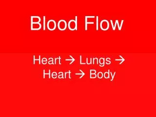
Fetal Circulation • No circulation to lungs • Foramen ovale • Ductus arteriosum • Circulation must go to placenta • Umbilical aa., vv.