Download
1 / 60
620 likes | 1.32k Views
The Peripheral Nervous System. The Peripheral Nervous System. Nervous structures outside the brain and spinal cord Nerves allow the CNS to receive information and take action Functional components of the PNS Sensory inputs and motor outputs Categorized as somatic or visceral

E N D
The Peripheral Nervous System • Nervous structures outside the brain and spinal cord • Nerves allow the CNS to receive information and take action • Functional components of the PNS • Sensory inputs and motor outputs • Categorized as somatic or visceral • Sensory inputs also classified as general or special
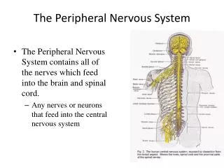
Basic Structural Components of the PNS • Sensory receptors – pick up stimuli from inside or outside the body • Motor endings – axon terminals of motor neurons • Innervate effectors (muscle fibers and glands) • Nerves and ganglia • Nerves – bundles of peripheral axons • Ganglia – clusters of peripheral neuronal cell bodies
Peripheral Endings • Afferent: Sensory Receptors • Efferent: Somatic Motor • Efferent: Autonomic Nervous System
Peripheral Sensory Receptors Afferent: Sensory • Structures that pick up sensory stimuli • Initiate signals in sensory axons
Peripheral Sensory Receptors • Two main categories of sensory receptors • Special nerve endings of sensory neurons • Monitor general sensory information • Independent receptor cells – specialized epithelial cells or small neurons • Monitor most types of special sensory information
Peripheral Sensory Receptors • Sensory receptors also classified according to: • Location • Type of stimulus detected • Structure
Classification by Location • Exteroceptors – sensitive to stimuli arising from outside the body • Located at or near body surfaces • Include receptors for touch, pressure, pain, and temperature • Interoceptors – (visceroceptors) receive stimuli from internal viscera • Monitor a variety of stimuli • Proprioceptors – monitor degree of stretch • Located in musculoskeletal organs
Classification by Modality • Mechanoreceptors – respond to mechanical forces • Thermoreceptors – respond to temperature changes • Chemoreceptors – respond to chemicals in solution • Photoreceptors – respond to light – located in the eye • Nociceptors – respond to harmful stimuli that result in pain
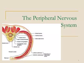
Classification by Structure • General sensory receptors • Widely distributed • Nerve endings of sensory neurons monitor: • Touch, pressure, vibration, stretch • Pain, temperature, proprioception • Divided into two groups • Free nerve endings • Encapsulated nerve endings
Free Nerve Endings • Abundant in epithelia and underlying connective tissue • Respond to pain and temperature • Monitor affective senses • Two specialized types of free nerve endings • Merkel discs – lie in the epidermis • Slowly adapting receptors for light touch • Hair follicle receptors – wrap around hair follicles • Rapidly adapting receptors
Encapsulated Nerve Endings • Consist of one or more end fibers of sensory neurons • Enclosed in connective tissue • Mechanoreceptors • Include four main types
Encapsulated Nerve Endings • Encapsulated nerve endings: dendrites with special supporting structures (mechanoreceptors and proprioceptors)
Encapsulated Nerve Endings • Meissner’s corpuscles • Pacinian corpuscles • Ruffini’s corpuscles • Proprioceptors
Proprioceptors • Monitor stretch in locomotory organs • Three types of proprioceptors
Three Types of Proprioceptors • Muscle spindles – measure the changing length of a muscle • Imbedded in the perimysium between muscle fascicles • Golgi tendon organs – located near the muscle-tendon junction • Monitor tension within tendons • Joint kinesthetic receptors • Sensory nerve endings within the joint capsules
Innervation of Skeletal Muscle • Motor axons innervate skeletal muscles • Neuromuscular junctions (motor end plates) • Similar to synapses between neurons • Acetylcholine diffuses across the synaptic cleft • Binds with molecules on the sarcolemma • Motor axons branch to innervate muscle fibers
Innervation of Skeletal Muscle • Motor unit – a motor neuron and all the muscle fibers it innervates
Innervation of Visceral Muscle and Glands • Simpler than neuromuscular junctions of skeletal muscle • Near the smooth muscle or gland it innervates • Visceral motor axon swells into a row of varicosities • Visceral motor responses • Slower than somatic motor reflexes
Cranial Nerves • Attach to the brain and pass through foramina of the skull • Numbered from I–XII • Cranial nerves I and II attach to the forebrain • All others attach to the brain stem • Primarily serve head and neck structures • The vagus nerve (X) extends into the abdomen
CN I: Olfactory Nerves • Sensory nerves of smell • Sense of smell • Damage causes impaired sense of smell
CN II: Optic Nerve • Sensory nerve of vision • Provides vision • Damage causes blindness in visual field
CN III: Oculomotor Nerve • Innervates four of the extrinsic eye muscles • Somatic and Autonomic motor function • Eye movement (Superior, inferior, medial rectus muscles and inferior oblique muscle), opening of eyelid (levator palpebrae superioris), constriction of pupil (circular muscle), focusing (ciliary muscle and accomodation) • Damage causes drooping eyelid, dilated pupil, double vision, difficulty focusing and inability to move eye in certain directions
CN IV: Trochlear Nerve • Innervates an extrinsic eye muscle • Eye movement (superior oblique muscle) • Damage causes double vision and inability to rotate eye inferolaterally
CN V: Trigeminal Nerve • Provides sensory innervation to the face • Motor innervation to chewing muscles • Ophthalmic branch – sensations from nasal cavity, skin of forehead, upper eyelid, eyebrow, nose • Maxillary branch – sensations from lower eyelid, upper lips and gums, teeth of the maxilla, cheek, nose, palate, pharynx • Mandibular branch – sensations from teeth of the mandible, lower gums and lips, palate, tongue. Motor function of temporalis and masseter muscles. • Damage produces loss of sensation and impaired chewing
CN VI: Abducens Nerve • Abducts the eyeball • Provides eye movement (lateral rectus m.) • Damage results in inability to rotate eye laterally and at rest eye rotates medially
CN VII: Facial Nerve • Innervates muscles of facial expression • Sensory innervation of face • Taste • Somatic Motor - facial expressions • Autonomic Motor - salivary and lacrimal glands, mucous membranes of nasal and palatine mucosa • Special Sensory - taste on anterior 2/3’s of tongue • Damage produces sagging facial muscles and disturbed sense of taste (no sweet and salty)
Branches of Facial Nerve Clinical test: Test anterior 2/3’s of tongue with substances such as sugar, salt, vinegar, and quinine; test response of tear glands to ammonia fumes; test motor functions by asking subject to close eyes, smile, whistle, frown, raise eyebrows, etc.
CN VIII: Vestibulocochlear Nerve • Sensory nerve of hearing and balance • Special Sensory • Provides hearing (cochlear branch) and sense of balance (vestibular branch) • Damage produces deafness, dizziness, nausea, loss of balance and nystagmus
CN IX: Glossopharyngeal Nerve • Sensory and motor innervation of structures of the tongue and pharynx • Taste • Somatic motor – Swallowing and voice production via pharyngeal muscles • Autonomic motor - salivation, gagging, control of BP and respiration • Sensations from posterior 1/3 of tongue including taste • Sensations from baroreceptors and chemoreceptors • Damage results in loss of bitter and sour taste and impaired swallowing, blood pressure anomalies (with CN X).
CN X: Vagus Nerve • A mixed sensory and motor nerve • Main parasympathetic nerve • “Wanders” into thorax and abdomen
Vagus Nerve X • Sensations from skin at back of ear, external acoustic meatus, part of tympanic membrane, larynx, trachea, espophagus, thoracic and abdominal viscera • Sensations from bararoceptors and chemoreceptors • Special sensory – taste from epiglottis and pharynx • Somatic motor – Swallowing and voice production via pharyngeal muscles • Autonomic motor – smooth muscle of abdominal viscera, visceral glands secretions, relaxation of airways, and normal or decreased heart rate. • Damage causes hoarseness or loss of voice, impaired swallowing, GI dysfunction, blood pressure anomalies (with CN IX), fatal if both are cut
CN XI: Accessory Nerve • An accessory part of the vagus nerve • Somatic motor function of pharynx, larynx, neck muscles • Swallowing, head, neck and shoulder movement via trapezius and sternocleidomastoid and pharyngeal muscles • Damage causes impaired head, neck, shoulder movement
CN XII: Hypoglossal Nerve • Runs inferior to the tongue • Innervates the tongue muscles • Tongue movements for speech, food manipulation and swallowing • If both are damaged – can’t protrude tongue • If one side is damaged – tongue deviates towards injured side
Cranial Nerve Disorders • Trigeminal neuralgia (tic douloureux) • recurring episodes of intense stabbing pain in trigeminal nerve area (near mouth or nose) • pain triggered by touch, drinking, washing face • treatment may require cutting nerve • Bell’s palsy • disorder of facial nerve causes paralysis of facial muscles on one side • may appear abruptly with full recovery within 3-5 weeks