Download
1 / 65
660 likes | 1.1k Views
Brain Imaging & the Mirror Neuron System. Lisa Aziz-Zadeh. Brain Imaging Methodologies. Fuctional Magnetic Resonance Imaging (fMRI) Transcranial Magnetic Stimulation (TMS). MRI and fMRI. MRI: Images of brain structure. fMRI: Images of brain function.

E N D
Brain Imaging & the Mirror Neuron System Lisa Aziz-Zadeh
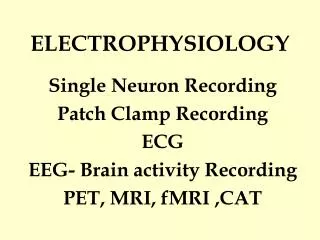
Brain Imaging Methodologies • Fuctional Magnetic Resonance Imaging (fMRI) • Transcranial Magnetic Stimulation (TMS)
MRI and fMRI • MRI: Images of brain structure. • fMRI: Images of brain function. • Tissues differ in magnetic susceptibility (grey matter, white matter, cerebrospinal fluid)
Physiological basis for Blood Oxygen Level Dependent(BOLD) fMRI • Neural activity leads to increased blood flow. • Increased flow exceeds increased oxygen extraction, resulting in decreased deoxy-hemoglobin content. • Deoxyhemoglobin is paramagnetic, so reducing the deoxy-Hb/oxy-Hb ratio increases the signal.
FMRI signal and neural activity • Recently it hasbeen shown thatthe BOLD signalreliably reflectsneural activity(Logothetis et al., 2001). • BOLD signal was correlatedwith both local field potentials(reflecting input) and multi-unit activity (reflecting output) • Note the lag of the BOLDresponse relative to theneural activity.
Signal Condition Time Acquiring functional images • Rapid sequence • Low resolution • Cognitivemanipulation • Statistics TASKREST
Statistical parameter maps • Every voxel has associated statistics. • SPMs are superimposed on anatomical images, thresholded and clustered.
Transcranial Magnetic Stimulation (TMS) as a Brain Mapping Tool
How TMS works • Pass a current through a hand held coil, whose shape determines the properties and the size of the field • The coil is driven by a machine which switches the large current necessary in a very precise and controlled way • The coil is held on the scalp and the magnetic field passes through the skull and into the brain • Small induced currents can then make brain areas below the coil more or less active, depending on the parameters used.
Safety Issues • Generally thought to be free from harmful effects • Examination of brain tissue submitted to thousands of TMS pulses has shown no detectable structural changes • It is possible in unusual circumstances to trigger a seizure in normal patients, but using the proper guidelines eliminate this risk
Different Types of TMS • Single Pulse • In the motor cortex, this usually causes a excitation in the brain. • rTMS • Multiple pulses in a short interval • Usually causes a “temporary lesion”
TMS as a Treatment Technique • Epilepsy • May be able to lower the number of seizures a patient • Depression • TMS treatments have been shown to cause improvement in severe cases of depression • An alternative to ECT
TMS to understand the motor system • Understanding the motor system • Participants watch different things that we think may activate the motor system • If these stimuli do activate the motor system, the participant’s muscles are just beneath the threshold of movement • We record the muscle activity when we give TMS over the motor cortex • If the stimulus had an effect, then we see bigger muscle twitches than if the stimulus had no effect
Using TMS with fMRI • fMRI -uses amount of blood flow used by the brain to determine which areas are the most active (more active areas use more oxygen)
fMRI • Problem: • are the areas shown to be used in an fMRI image ESSENTIAL to the brain function, or are do they activate peripherally? • ROLE OF TMS: • Using the “inhibitory lesion technique” we can turn off the specific brain area and see if it is ESSENTIAL for the task. If the person can not perform the task during rTMS, it is essential.
Cortical Mechanism forAction Recognition adds additional somatosensory information to the movement to be imitated provides an early description of the action Observed Action STS Parietal mirror neurons (PF) (inferior parietal lobule) Frontal mirror neurons (F5) (BA 44) copies of the motor plans necessary to imitate actions for monitoring purposes codes the goal of the action to be imitated
A B C Imitation of Hand Actions Iacoboni et al. 1999
Posterior Parietal Cortex Execution Observation + + Iacoboni et al. 1999
Observation Execution Superior Temporal Sulcus and Imitation
Summary • Anatomical similarity between human and nonhuman primate frontoparietal mirror systems • Broca’s area codes the goal of the action (lift the finger) • PPC codes the precise kinesthetic aspects of the movement (how much the finger should be lifted) • STS codes the visual information (input) • Both left and right hemispheres are active
Auditory Mirror Neurons peanut breaking squeekingduck vision & sound 100 spk/s 0 1s vision sound motor Christian Keysers
Acoustic Mirror Neurons in the Monkey: Kohler et al (2002) • Discriminated significantly between two different sounds of actions (ripping paper, breaking a peanut) • Representation of actions in these neurons are independent both of who performs the actions and how they are perceived • Multimodality may provide a first step towards abstract, semantic representations, perhaps tying to origin of language
Can we get a similar result in humans? A study using TMS
Transcranial Magnetic Stimulation (TMS) Study • Q: • Do acoustic mirror neurons exist in the human brain? • Is there hemispheric specialization for the auditory modality? • Single pulse TMS to left or right primary motor (M1) hand area • Motor Evoked Potentials (MEPs) recorded from the left or right hand muscle (FDI) • Subject listens to 3 auditory stimuli: • Bimanual Hand Action Sound: Typing orTearing Paper • Bipedal Leg Action Sound: Walking • Control Sound: Thunder
Predicted Results • Prediction: • MEPs will be largest when the action sound matches the muscles of the stimulation site • Left hemisphere specialization
Results: Significant Facilitation to Hand Stimuli in the Left Hemisphere * *
Discussion • Motor facilitation to action sounds • Left hemisphere specialization • All the components of an action seem to be available to left hemisphere
Mirror Neurons and understanding another person’s INTENTIONS
Mirror neurons respond to GOAL ORIENTED actions, even when only the intent is apparent but the action itself is occluded • May have implications for INTENTION UNDERSTANDING • fMRI studies in Humans show similar findings (Iacoboni et al, 2004)
Mirror Neurons and Language Language Evolution Embodied Semantics