Download
1 / 1
20 likes | 65 Views
Supplement 1. Parotid. WKY WKY-D SHR SHR-D. B. A. C. D. E. F. G. H.

E N D
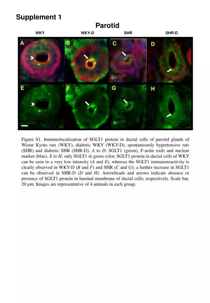
Supplement 1 Parotid WKY WKY-D SHR SHR-D B A C D E F G H Figure S1. Immunolocalization of SGLT1 protein in ductal cells of parotid glands of Wistar Kyoto rats (WKY), diabetic WKY (WKY-D), spontaneously hypertensive rats (SHR) and diabetic SHR (SHR-D). A to D: SGLT1 (green), F-actin (red) and nuclear marker (blue). E to H: only SGLT1 in green color. SGLT1 protein in ductal cells of WKY can be seen in a very low intensity (A and E), whereas the SGLT1 immunoreactivity is clearly observed in WKY-D (B and F) and SHR (C and G); a further increase in SGLT1 can be observed in SHR-D (D and H). Arrowheads and arrows indicate absence or presence of SGLT1 protein in luminal membrane of ductal cells; respectively. Scale bar, 20 µm. Images are representative of 4 animals in each group.