Download
1 / 38
390 likes | 586 Views
Tissues of the Body Module (TOB) Introductory Lecture LIGHT MICROSCOPY. Histology Textbooks ‘Basic Histology’, Junqueira , ‘ Colour Atlas of Histology’ Gartner and Hiatt.

E N D
Tissues of the Body Module(TOB)Introductory LectureLIGHT MICROSCOPY
Histology Textbooks‘Basic Histology’, Junqueira, ‘Colour Atlas of Histology’Gartner and Hiatt
Histologystudy of the structure of tissuesby means of special stainingtechniques combined with light andelectron microscopy.
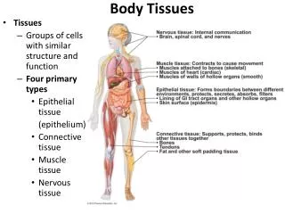
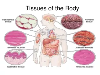
state the meaning of the term tissue Tissue – a collection of cells specialized to perform a particular function. Aggregations of tissues constitute organs.
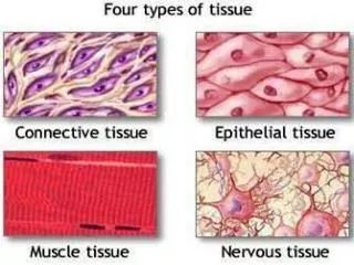
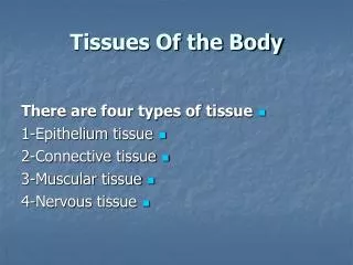
Tissue Classification1. Epithelial tissue2. Connective (Support) tissue3. Muscle tissue4. Nervous tissue
The relationship between milli-, micro and nanometers.meter mmillimeter mm 10-3mmicrometer µm 10-6 mnanometer nm 10-9 mAngstrom Unit Å 10-10m
Most human cells are 10 – 20 µm indiameter (about 5 times smaller thanthe smallest visible particle).
Biopsy – the removal of a small pieceof tissue from an organ or part of thebody for microscopic examination.
Types of biopsySmear – e.g. cervix Curettage – e.g. endometrial lining of uterusNeedle – e.g. brain, breast, liver, kidney, muscleDirect incision – e.g. skin, mouth, larynxEndoscopic – e.g. lung, intestine, bladderTransvascular– e.g. heart, liver
why tissue needs to be fixed andwhich fixativesare commonly used.
Fixation confers stability upon tissue. Unfixed tissue is subject to attack by bacteria (putrefaction) and by the enzymes that are present within the cells themselves (autolytic enzymes). Fixation is directed primarily towards the preservation of proteins by making them insoluble. Formaldehyde and glutaraldehyde are commonly used as fixatives. These reactive aldehydes form covalent bonds with the free amino groups of proteins and thus cross-link adjacent proteins, arresting biological activity and making cells more amenable to staining.
Tissue Processing Procedure 1-Fixation2-Dehydration and clearing3- Wax embedding4- Cutting and Mounting section 5- Staining 6- Mounting
A microscope is an instrument for viewing objects that are too small to be seen by our naked eyes. The definition of microscopic means minute or very small, not visible with the eye unless aided by microscope
Types of the microscope There are many types of microscopes , ranging from simple , single – lens instruments ( magnifying glasses ) to compound microscope and high- powered electron . two basic types of microscopes that are used in biological studies : the compound light microscope and the electronmicroscope
Magnification Your microscope has 3 magnifications : Scanning , Low and High . each objective will have written the magnification . In addition to this , the ocular lens (eye piece ) has a magnification . The total magnification is the Ocular × Objective .
electron microscope is a type of microscope that uses a beam of electrons to illuminate the specimen and produce a magnified image. 1- Scanning electron microscope 2- transmission electron microscope
Transmission electron microscope (TEM)The original form of electron microscope, the transmission electron microscope (TEM) uses a high voltageelectron beam to create an image.
Scanning electron microscopeUnlike the TEM, where electrons of the high voltage beam carry the image of the specimen, the electron beam of the scanning electron microscope (SEM )does not at any time carry a complete image of the specimen. The SEM produces images by probing the specimen with a focused electron beam that is scanned across a rectangular area of the specimen.