Download
1 / 64
640 likes | 839 Views
The Digestive System. Subdivisions of the Abdomen. Commonly used abdominal incisions: Any incisions or scars present? Presence of an incision may speculate what the surgeon might have done.

E N D
Commonly used abdominal incisions: • Any incisions or scars present? Presence of an incision may speculate what the surgeon might have done. • Subcostal incision on the right---------------------open bladder surgery (cholecystectomy). • Barely visible round scars around the umbilicus may suggest closed or laparoscopic cholecystectomy • Left subcostal incision----------------to approach the spleen • Suprapubic incisions is used to access pelvic organs----------------(C-section,hysterectomy) • An oblique incision approximately at the meeting point of umbilical region and ® inguinal region is frequently used for appendeceptomy. • A scar in one or both inguinal region may be indicative of previous hernia surgery.
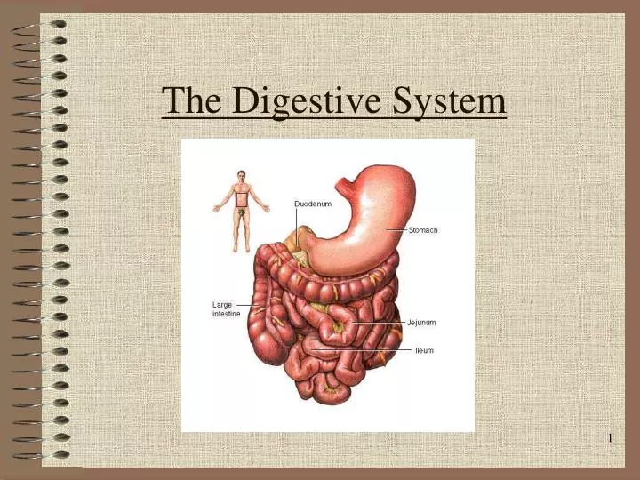
Digestive sytem: • -The organs that are involved in breaking down of food into molecules that can pass through the walls of the digestive tract and can be taken up by the cells. • Digestion process are in two stages: • - mechanical digestion • Chemical digestion • G I tract or Alimentary canal: • A continuous tube the begins at the mouth and ends at the anus.
Digestion • Processing of food • -2 Types of processing: • Mechanical (physical) • Chew • Tear • Grind • Mash • Mix • Chemical • Catabolic reactions • Enzymatic hydrolysis • Carbohydrate • Protein • Lipid
Digestion • Phases • Ingestion • Movement • Digestion • Absorption • Further digestion **chyme partially digested food(a thick, heavy,creamlike liquid).***
Digestive System Organization • Gastrointestinal (Gl) tract (Alimentary canal) • Tube within a tube • Direct link/path between organs • Structures • Mouth • Oral Cavity • Pharynx • Esophagus • Stomach • Duodenum • Jejenum • Ileum • Cecum • Ascending colon • Transverse colon
Descending colon Sigmoid colon Rectum Anus Accessory structures Not in tube path Organs Teeth Tongue Salivary glands Liver Gall bladder Pancreas Digestive System Organization
Figure 15—2. Surface of the tongue on the region close to its V-shaped boundary, between the anterior and posterior portions. Note the lymphoid nodules (lingual tonsil), glands, and papillae.
Sequence: Voluntary stage Push food to back of mouth Pharyngeal stage Raise Soft palate Larynx + hyoid Tongue to soft palate Esophageal stage Contract pharyngeal muscles Open esophagus Start peristalsis Deglutition (swallowing)
Control Nerves Glossopharyngeal Vagus Accessory Brain stem Deglutition center Medulla oblongata Pons Disorders Dysphagia Aphagia Deglutition (swallowing)
Esophagus • Surrounded by • SNS plexus • Blood vessels • Functions • Secrete mucous • Transport food
2 types of muscular action occur in the small intestines: **Segmentation and peristalsis.** Segmentation is the muscle action that mixes chyme and digestive juices, like a cement mixer. The second action is peristalsis, moving undigested food remains toward the large intestine
Esophagus • Sphincters: • Upper (pharyngoesophageal sphincter) • Lower (LES) • Abnormalities ??
Stomach • Usually “J” shaped • Left side, anterior to the spleen • Mucous membrane • G cells – make gastrin • Goblet cells – make mucous • Gastric pit – Oxyntic gland – Parietal cells – Make HCl • Chief cells – Zymogenic cells • Pepsin • Gastric lipase
3 muscle layers Oblique Circular Longitudinal Regions Cardiac sphincter Fundus Antrum (pylorus) Pyloric sphincter Vascular Inner surface thrown into folds – Rugae Contains enzymes that work best at pH 1-2 Stomach
Functions Mix food Reservoir Start digestion of Protein Nucleic acids Activates some enzymes Destroy some bacteria Makes intrinsic factor – B 12 absorption. The stomach’s activity is controlled by the PNS*** Absorbs Alcohol Water Stomach
1.The deep folds of the stomach wall that allow for size changes of the stomach are called? • Rugby • Sphincter • Rugae • Glottal folds • 2- the stomach’s activity is controlled by the _______ nervous system. • 3- the final “door” of the stomach that needs to open for chyme to travel to the small intestine is located at the end of the ? • Fundus f. ???? • Epiglottis • Adventitia • Serosa • Cardiac region
Extends from pyloric sphincter ileocecal valve Regions Duodenum Jejunum Ileum Movements Segmentation Peristalsis Small Intestine
The small intestines is small in diametre,not in length. Beginning from the pyloric sphincter,the small intestines is also the longest section of the alimentary canal ,with length of up to 20 feet(up to 6 metres). In small intestine, almost 80% of the absorption of usable nutrients takes place when chyme comes in contact with the mucosal walls. Simple sugars, Aa, fatty acids, vitamins and water are all absorbed here.***** Some of the remaining 20% was already absorbed in the stomach, with the rest being absorbed in the large intestines. Any residue that cannot be utilised is sent on to the large intestine for removal from the body as fecal matter(feces).
Small Intestine • Histology features • Intestinal glands – Intestinal enzymes • Duodenal glands – Alkaline mucous • Paneth cells – Lysozyme • Microvilli • Lacteals • Plica circularis • Smooth muscle • Lymphatic tissue – GALT • Vascular
Absorbs 80% ingested water Electrolytes Vitamins Minerals Carbonates Active/facilitated transport Monosaccharides Proteins Di-/tripeptides Amino acids Lipids Monoglycerides Fatty acids Micelles Chylomicrons Small Intestine
The structure of the wall of the small intestine possesses circular folds called plicae circulares and finger-like protrusions into the lumen called villi. The villi which posses microscopic extensions known as microvilli. These villi are tightly packed, giving the mucosa velvety texture and appearance. The purpose of the microvilli ,villi and circular folds is to provide an incredible increase in the surface area of the small intestine. Each villus contains a network of capillaries and a lymphatic capillary called LACTEAL. Intestinal glands are located between villi. The capillaries absorb and transport sugars the result of carbohydrate digestion) and amino acids (the result of protein digestion) to the liver for further processing before they are sent throughout the body.
Glycerol and fatty acids(obtained from the digestion of fat), are absorbed by the villi and converted into a lipoprotein that travels on to the lacteal, where it is now a white, milky substance called chyle. Chyle goes directly into the blood stream via the left subclavian vein for distribution throughout the body. The walls of the small intestine secrete several enzymes important for the final stages of chemical digestion and two hormones that control the activity of the pancreas, gallbladder and stomach. The pancreas is stimulated to secrete as a result of the hormone secretin that is produced by the small intestines. gall bladder activity is stimulated by the hormone cholecystokinin,which is produced by the small intestines.
At the duodenum, additional secretions are added from the pancreas and gall bladder. The pancreas provides pancreatic juices, and the gall bladder produces bile. Bile emulsifies fat, (that is it makes fat able to disperse in water making the fat easier to breakdown.) Pancreatic juice contains enzymes and sodium bicarbonate, which neutralises the acidic chyme. The small intestine also produces digestive enzymes that are needed to complete chemical digestion. These enzymes and (mucus) are produced by exocrine cells. lactase, maltase,and sucrase are needed for the digestion of double sugars such as disaccharides that are contained in starches. Peptidases--- needed to digest portions of the protein structure called peptides. Intestinal lipase—for digestion of certain fats.
Hormone secreting organ action Gastrin stomach stimulates release gj. Secretin duodenum stimulates release of bicarbonate and water from pancreas and bile from liver. Cck duodenum stimulates digestive enzyme release from the pancreas and bile release from the gall bladder. *** it is important to note that the secretion of these substances is mainly due to the presence of chyme in the small intestines.****
Secretes digestive enzymes Peptidases Amino- Di- Tri- Sucrases Maltase Lactase Saccharidases Di- Tri- Lipase Nucleases Small Intestine
Control Requires pancreatic enzymes & bile to complete digestion Small Intestine
Large Intestine • Extends from ileocecal valve to anus • Regions • Cecum – Appendix • Colon • Ascending • Transverse • Descending • Rectum • Anal canal
The large intestines is responsible for : • -water reabsorption • -absorption of vitamins produced by normal bacteria in the large intestines • Packaging and compacting waste products for elimination from the body. • ** because there are no villi in the large intestines, little nutient absorption occurs here.*** • Approximately 1.5metres long 7.5 cm in diametre, large intestine is divided into 3 main regions; the cecum, colon, and rectum. • A pouch-shaped structure, the cecum recieves any undigested food ,such as cellulose and water from the ileum of the S.I. • The infamous appendix is attached to the cecum, about 9cm long, the appendix is a slender, hollow dead-end tube lined with lymphatic tissue.
Because it possesses lymphatic tissue it helps to fight infection ,and also discovered to replenish the beneficial bacteria in our digestive tract. Appendicitis?? Peristalsis continues in the large intestine but at a slower rate. As these slower, intermittent waves move fecal matter toward the rectum, water is removed, turning the feces from a watery soup to a semisolid mass. Some of the water (used in digestion) and electrolytes are reabsorbed by the cecum and asc. Colon. Although this is a relatively small amount of water reabsorbed, it is crucial in maintaining the proper fluid balance in the body. As the rectum fills up with feces,a defecation reflex occurs, which causes rectal muscles to contract and anal muscles to relax.
Defecation is controlled by a combination of voluntary and Involuntary reflexes. 2 sphincters sorround the anal opening, IAS AND EAS. When feces enters the rectum the wall stretches, triggering the defecation reflex. Diarrhea ----- if fecal matter moves through the large intestines too rapidly, not enough water is adequately reabsorbed. Constipation----- if the fecal matter remains too long in the large intestines, too much water is removed. Feces hardens. Functions of the large intestines: I bacteria aids further break down of indigestible material. II produce B- complex vitamins III produces most of the vitamin k ****bacteria outside the intestinal wall in the blood stream, could be fatal***
Large Intestine • Histology • No villi • No permanent circular folds • Smooth muscle • Taeniae coli • Haustra • Epiploic appendages • Otherwise like rest of Gl tract
Large Intestine • Functions • Mechanical digestion • Haustral churning • Peristalsis • Chemical digestion – Bacterial digestion • Ferment carbohydrates • Protein/amino acid breakdown • Absorbs • More water • Vitamins • B • K • Concentrate/eliminate wastes
Chyme dehydrated to form feces Feces composition Water Inorganic salts Epithelial cells Bacteria Byproducts of digestion Defecation Peristalsis pushes feces into rectum Rectal walls stretch Control Parasympathetic Voluntary Feces Formation and Defecation
Liver • Location • R. Hypochondrium • Epigastric region • 4 Lobes • Left • Quadrate • Caudate • Right • Each lobe has lobules – Contains hepatocytes – Surround sinusoids – Feed into central vein • Weighing approx.1.5kg(3.3 pounds).
Functions Makes bile Detergent – emulsifies fats Release promoted by: Vagus n. CCK Secretin Contains Water Bile salts Bile pigments Electrolytes Cholesterol Lecithin Liver
Detoxifies/removes Drugs Alcohol Stores Glycogen Vitamins (A, D, E, K) Fe and other minerals Cholesterol Activates vitamin D Fetal RBC production Phagocytosis Metabolizes absorbed food molecules Carbohydrates Proteins Lipids Liver
Dual blood supply Hepatic portal vein Direct input from small intestine Hepatic artery/vein Direct links to heart Liver