Download
1 / 18
210 likes | 394 Views
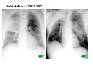
Explore radiological aspects of pneumonia, pulmonary emphysema, and interstitial pneumonia, along with bronchiectasis, atelectasis, and chronic pulmonary abscess. Detailed insights on various pathologies, abscesses, and cancers are provided in this informative guide.

E N D
TRIANGLE DITTMAR RUPERT INTERSTITIAL PNEUMONIA
- The same case; mixed image with horizontal line (air-fluid)
Bronchiectasis- triangle BILATERAL BRONCHIECTASIS: suprainfection
Bronchial patology ULCERATIONS
CHRONIC PULMONARY ABSCESS
BRONCHOPULMONARY CANCER Left hilary opacity;
-ATELECTASIS right lung atelectasis - volume reduction , attraction of mediastinum, diaphragm, rib grid
1 2 Total pneumothorax 1 Hyper transparency with no vascular design 2 - collapsed lung to hilum Total left pneumothorax
X-ray: opacity of homogeneous intensity that varies by the amount of liquid Typical image of right basal pleurisy – moderate intensity of the opacity, with the upper limit, flou, in meniscus, upper concave
homogeneous opacity right hemithorax, controlateral mediastinal shift