Download
1 / 53
710 likes | 1.66k Views
Acute Necrotizing Pancreatitis. Yoram Klein MD. MAGNITUDE OF THE PROBLEM. The disease may be mild and self limiting, 70-80% take course of edematous interstitial inflammation Necrotizing pancreatitis develops in 20-25% pts . 20-30% will develop local or systemic complications

E N D
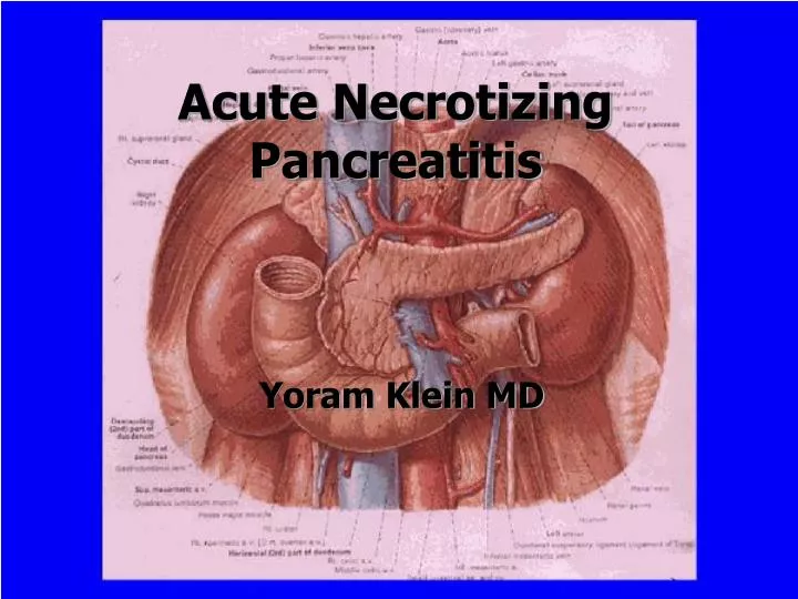
Acute Necrotizing Pancreatitis Yoram Klein MD
MAGNITUDE OF THE PROBLEM • The disease may be mild and self limiting, 70-80% take course of edematous interstitial inflammation • Necrotizing pancreatitis develops in 20-25% pts . • 20-30% will develop local or systemic complications • Approx 1 in 4 pts who develop complications will die
AP & QUESTIONS • WHAT IS THE CORRCT DIAGNOSIS? • What is the prognosis? • Are complications developing? • Can an associated condition to be identified? • What is the ideal timing for surgery?
OBJECTIVE • To give pts of AP best chance of survival, from the outset to be managed by surgeon • Identification of pts likely to develop complications • Management (prevention)of systemic complications • Timing and choice for surgical Intervention for gall stones or local complications
PANCREATITIS (terminology) • MILD-uncomplicated recovery • SEVERE-AP with evidenceoffailure of one or more systems ,or local complication. • These terms are defined retrospectively,when outcome is known • Prospectively defined on the basis of scoring systems.Predicted Mildor Predicted Severe
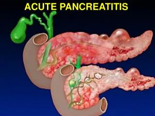
ACUTE PANCREATITITS-TERMINOLOGY • COMPLICATED-local or systemic complications • EDEMATOUS-Swollen, red ,with or without fat necrosis;Histology fluid,debris,leukocytes present • PERIPANCREATIC NECROSIS-Necrosis of retroperitoneal fat, other organs rarely involved, occasionally infarction by vascular thrombosis.This change may be present alone or may coexist with or be absent in presence of pancreatic necrosis
AP-local complications ……contd • Pancreatic necrosis; • Patchy or diffuse superficial or parenchymal necrosis, unequivocally demonstrated by inspection after opening of the pancreatic capsule , or histological criteria; local or diffuse areas of non enhancement on CT, sterile necrosis • Infected pancreatic necrosis; Necrosis with positive bacterial cultures • Pancreatic abscess;Loculated walled off collections of pus as a late complication of AP, usually after 3 weeks
MANIFESTATIONS OF AP • LOCAL; • MILD; • EDEMA, INFLAMMATION, NECROSIS • SEVERE; • PHLEGMON, NECROSIS, INFECTION, FLUID COLLECTION, ABSCESS
Bacterial contamination • Risk of bacterial infection on necrotic tissue • 60% in proven cases of NP • Risk in ist week =25% • Risk in 2nd week = 35-40% • Risk in 3rd week =60% • Organisms are Gram negative E-coli,Proteus,Pseudomonas,staphylococci
SYSTEMIC COMPLICATIONS • Respiratory-Interstitial pulmonary edema;gas transfer impairment,Pt may need ventilation • Renal-oliguria-require aggressive circulatory support,#Dialysis • Cardiovascular-Hypotension, edema,aggressive fluid therapy and Ionotropes • Haemopoiesis, Coagulation system, Endocrine systems
PANCREATITIS • How to diagnose it? • How to evaluate severity? • RANSON CRITERIA • IMRIES CRITERIA • APACHE scoring • GLASGOW Criteria • Lab and Radiology Help ;
Diagnosis of Pancreatitis • Clinical Diagnosis • Lab studies; • Serum amylase;Levels Rise within 2-12hrs, • 3x times normal is cut off . (n35-118 IU/liter • levels normal in 2-3days. • Persistence of ^ levels >10days denote complication like cyst,abscess. • 5%cases no increase value
Diagnosis of pancreatitis(contd) • Serum lipase ^^ 2x times the normal( 2.3-20.0 IU/L) n=3-5days • CR protein,LDH ,Serum Neutrophil –elastase,IL-6, and alpha macroglobulin • Trypsin like Immunoreactivity
Initial 24 hrs 1.Age >55 years 2.Glucose >than 200 mgm/dl 3.WBC > 16,000 cells/mic L 4.LDH >350 IU/liter 5.AST >250IU/liter Subsequent 48 hrs 1.Art o2tension <60mmHg 2.Bun Increase >8mg/dl 3.Ca < 8mg/dl 4.Base deficit >4meq/liter 5.Estimated fluid sequestration >6liters 6.Fall n Hct >10% RANSON CRITERIA
Mortality prediction (as per Ranson criteria) • A. < 3 signs = 1% • B. Three to Four signs=11% • C. Five to six signs=33% • D. >Six signs= 100%
Temp Mean Art Pressure Heart Rate Resp rate Oxygenation(Pao2) Arterial Ph Serum sodium SerumPottasium Serum creatinine Haematocrit WCC Glasgow coma scale APACHEII
A=+4 to 0 points TEMP>41=4,<29=4 Mean Art Pr>160=4 <49=4 Heart & Resp rateOXYGENATIONART PHSer Na,K,Creat, HCT,WBC GLASGOW COMA Score B=Age <44=0 pts >75=6points C=Chronic Health points H/o organ insufficiency Liver,CVS,Resp,Renal, ,Immunocompromised APACHE SCORE42=90% Mort Apache II score(Sum of A+B+C)
GLASGOW CRITERIA • Any time during First 48hrs after admission • 1.WBC >15000 Cu/mm • 2.Blood glucose>10mmol/l • 3.BUN >16mmol/L • 4.Art po2,< 60mmHg • 5.Ser ca. <2.0 ml/l • 6.Ser Albumin<32gm/l • 7.Ser LDH >600u/L(n=250) • 8.AST Or ALT >200u/l
Any time during First 48hrs after admission; WBC >15000 Cu/mm Blood glucose>10mmol/l BUN >16mmol/L Art po2,< 60mmHg Ser ca. <2.0 ml/l Ser Albumin<32gm/l Ser LDH >600u/L(n=250) AST Or ALT >200u/l GLASGOW CRITERIA
INTERSTITIAL AND NECROTIZING PANCREATITIS (Discrimination) • Markers of Necroses • C-reactive protein>120 mgm/L • PMN-Elastase>120mgm/L • PLA>15U/L • PLA2>3.5U/L • Dynamic angio –CT • Guided needle aspiration of necroses for detection of bacteria
RADIOLOGY • Plain Films • Ultrasonography • Sens;62-95%,Specif>95%, pancreas not visualized in> 40%pts • CT scan;Sens 90% Specif+100% • ERCP • PTC. Pancreatitis is due to gallstone? Or Alcoholic?
Enlargement of Gland Ill defined margins Abnormal enhancement Thickening of peripancreatic planes Blurring of fat planes Intra & retroperitoneal fluid collection Pleural effusion Pancreatic gas indicative of necrosis /abscess Pseudocyst formation CT findings in Acute Pancreatitis
ERCP; Indications In AP • Preop evaluation with suspected traumatic pancreatitis to see Pancreatic duct disruption • Pts with suspected biliary Pancreatitis and severe disease and not clinically improving by 24hrs after admission. Do ERCP for stone extraction
ERCP-indications (contd • In pts >40 with no identifiable disease to rule out occult CBD stones,pancreatic or ampullary Ca or other causes of obstruction; • Pts <40 at a post Cholecystectomy status or more than one attacks of unexplained pancreatitis
SYSTEMIC TREATMENTS • Basic principles-ICU,Rest GIT and Pancreas,analgesia,oxygenation • Pancreatic inhibition (Glucagon, Somatostatin)? • Antibiotics • Nutrition (Enteral route is safe& preferred )
Role of Antibiotics in AP • Traditional teaching Prophylactic antibiotics do not prevent abscess- • Mezlocillin, Metrionidazole, Imipnem good concentration in pancreatic juice • Cefotaxime, Ceftazidime Clindamycin, Ciprofloacin good levels in p. juice • They can limit rate of infection of this necr material(Bossi1992)
Operative Measures For AP • A.Diagnostic laparotomy • B.To limit the severity of pancreatic inflammation • Biliary operations • C.To interrupt the pathogenesis of complications • Pancreatic drainage • Pancreatic resection • Peritoneal drainage
Operative measures(contg) • D.To support the patient and treat complications • Drainage of pancreatic abscesses • Feeding jejunostomy • To prevent recurrent pancreatitis
Diagnostic uncertainty Gall stone induced pancreatitis Pancreatic drainage and defunctioning Pancreatic resection Peritoneal Lavage Operation for complications Surgical treatment-indications
Bile duct stones-strategy • Acosta (1974), recovered gall stones from Faeces of pts with gall stone pancreatitis. • Neptolemos (1989) ;Passage of stone through ampulla precipitates pancreatitis attack, persistence of stones in CBD; Pt is at risk of complications and death • Early surgery or to deal with CBD stones endoscopically (ERCP) 14 %pts of AP have coexisting cholangitis
Timing OF Operation IN Gall Stone Pancreatitis • Mild pancreatitis:Operated At Any Stage during first admission • Severe disease.Cholecystectomy during first admission, timing depends on clinical indicators
Timing of Surgery-contd • RECOVERING PT.Allow pt to settle completely before elective early operation is taken prior to discharge. • UNSTABLE PT- Who will require surgery to deal with local complications of pancreas, Cholecystectomy to be performed at this time • Early Cholecystectomy within 48-72 hours of admission is best avoided in these all patients
Clinical criteria Surgical acute abdomen Sepsis syndrome Shock syndrome Non response to ICU Morphologic +Bacteriologic Infected necroses Extended pancreatic necrosis>50% Extnd. intrapancreatic +retroperitoneal necroses Indications of Operation IN NP
Technique of Debridement • Closed cavity Lavage • Open abdomen • Surgical drainage • Posterior approach • Pancreatic resection