anti titin antibody
110 likes | 126 Views
anti-Titin-Antibody ELISA Kit can be provided from Creative Diagnostics.t<br>https://www.creative-diagnostics.com/TTN-EIA-Kit-257559-471.htm<br>

anti titin antibody
E N D
Presentation Transcript
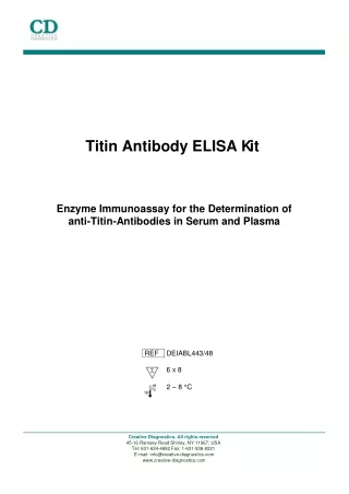
Titin Antibody ELISA K it Enzyme Immunoassay for the Determination of anti-Titin-Antibodies in Serum and Plasma REF DEIABL443/48 6 x 8 2 – 8 °C Creative Diagnostics. All rights reserved 45-16 Ramsey Road Shirley, NY 11967, USA Tel: 631-624-4882 Fax: 1-631-938-8221 E-mail: info@creative-diagnostics.com www.creative-diagnostics.com
Contents 1. Introduction and Principle of the Test 2. Precautions 3. Storage and Stability 4. Contents of the Test 5. Preparation of Reagents and Samples 6. Assay Procedure 7. Calculation and Interpretation of Results 8. Literature Page Page Page Page Page Page Page Page 11 3 4 4 4 6 7 8 Page 2
1. Introduction and Principle of the Test In about 80 % of patients with Myasthenia gravis alterations of the thymus can be detected. Approximately 10 % of these patients develop thymus neoplasia as thymic epithelial tumor (TET) or thymic carcinoma. An early diagnosis of the thymoma and subsequent thymectomy is decisive for the prognosis of these patients. Most of the affected patients develop acetylcholine receptor antibodies which can be detected with the ACHRAB-Assay for the diagnosis of myasthenia gravis, as well as autoantibodies against striated muscles, among others antibodies to titin. Titin is a protein of the striated muscles with an extremely high molecular weight. The immunogenic region of titin is located on a 30 kD protein fragment. Antibodies against this fragment presumably crossreact with the epitopes of the acetylcholine receptors (paraneoplastic myasthenia gravis). The recombinant MGT30 peptide is used in the ELISA for the specific determination of anti-titin antibodies. The new ELISA assay is clearly superior to the immunofluorescence test on sections of striated human or monkey muscles. The Anti-Titin ELISA kit uses the microtitre plate format. Recombinant titin fragment (MGT30 peptide) is coated onto the surface of the microwells. Diluted serum specimens are incubated to allow antibodies to titin to bind to the plastic surface. After washing away unbound antibodies and serum constituents, the specific titin antibodies are detected by protein A- peroxidase. The TMB / peroxidase reaction is monitored at 450 nm. Page 3
2. Precautions For in vitro research use only. Do not eat, drink or smoke where immunodiagnostic materials are being handled. Do not pipette by mouth. Some reagents contain sodium acid as preservative. Avoid skin contact. • Wear disposable gloves when handling immunodiagnostic material. Some kit components are made with human sera. All sera used were tested for HIV I/II antibodies, HCV and HBsAg and found to be negative. However, because no test method can offer complete assurance that infectious agents are absent, these reagents should be handled as potential biohazardous material. Material of animal origin used in the preparation of the kit has been obtained from animals certified as healthy but these materials should be handled as potentially infectious. 3. Storage and Stability On arrival, store the kit at 2-8 °C. Once opened the kit is stable until its expiry date. For stability of prepared reagents refer to Preparation of Reagents. Allow all reagents to reach room temperature before use. 4. Contents of the Test 4.1 MT-Strips STRIPS 8 wells each, break apart precoated with recombinant MGT30 peptide 4.2 Enzyme Conjugate CONJ 6 ml, ready for use Protein-A-POD conjugate 4.3 Calibrator CAL 1 ml serum, ready for use (1:101 prediluted) 4.4 Negative Control CON - 1 ml serum, ready for use (1:101 prediluted) 4.5 Positive Control CON + 1 ml serum, ready for use (1:101 prediluted) 6 strips 1 vial 1 vial 1 vial 1 vial Page 4
4.6 Sample Diluent DIL 55 ml, ready for use 4.7 Wash Buffer WASH 50 ml, concentrated Dilute with dist. water to 500 ml. 4.8 Substrate SUB 6 ml TMB solution, ready for use 4.9 Stop Solution STOPP 6 ml, ready for use Contains 0.3M sulphuric acid, not corrosive Reagents and materials required but not provided: Pipettes (20 µl, 100 µl, 1 ml) Repeating dispenser 100 µl Horizontal shaker Microplate washing device Microplate photometer Distilled water 1 bottle 1 vial 1 vial 1 vial Page 5
5. Preparation of Reagents and Samples Allow all reagents and required number of MT strips to reach room temperature. 5.1 Specimen Fresh plasma or serum samples are suitable. Samples should be stored at -20 °C if necessary. Repeated freezing and thawing, however, can affect the results. Hemolytic samples should not be used. Samples must be diluted 1 : 101 (e.g. 10 µl serum plus 1 ml diluent) in Sample Diluent before assay. Diluted samples should be stored frozen for repeated measurement. 5.2 MT-Strips STRIPS Keep the closed bag at room temperature for around 10 minutes. Take the strips that are not needed out of the frame and store them in the thoroughly closed bag (leave the desiccant in the bag). 5.3 Wash Buffer WASH Prepare the Wash Buffer by diluting with distilled water to a final volume of 500 ml. Diluted Wash Buffer is stable for two months at 2 - 8 °C. All other reagents are ready for use. Page 6
6. Assay Procedure 6.1 Sample Incubation Dispense 100 µl of each ready for use Calibrator, ready for use Negative and Positive Control and diluted patient samples into the corresponding wells. Incubate 60 minutes at room temperature on a horizontal shaker. 6.2 Washing Discard or aspirate the contents of the wells and wash thoroughly with each 300 µl Wash Buffer. Repeat the washing procedure 2 to 3 times. Remove residual liquid by tapping the inverted plate on clean absorbent paper. 6.3 Conjugate Incubation Dispense 100 µl Enzyme Conjugate into each well. Incubate for 30 minutes at room temperature on a shaker. 6.4 Washing Repeat the washing procedure as described in 6.2. 6.5 Substrate Incubation Dispense each 100 µl Substrate in the wells and incubate for 15 to 25 minutes on a shaker. 6.6 Stopping Dispense each 100 µl Stop Solution into the wells in the same order sequence as the Substrate. 6.7 Measurement Read the optical density at 450 nm (reference wavelength between 570 and 650 nm) in a microplate photometer within 10 minutes. Samples with an OD higher than 2.5 at 450nm should be read again at 405nm and should be evaluated against the calibrator read at 405 nm. Page 7
7. Calculation and Interpretation of Results The measured optical density of the samples divided by the optical density of the Calibrator equals factor F: O.D. Sample Factor Sample O.D. Calibrator Typical example: O.D. O.D. Interpretation Sample Calibrator 1.0 0.2 2.8 0.5 2.0 O.D.Sample Calibrator Negative Control Positive Control Sample 1 Sample 2 0.766 0.126 2.157 0.391 1.498 – + – + Samples with an OD higher than 2.5 at 450nm should be read again at 405nm and should be related to the calibrator read at 405nm. In the following table the results of the measurement of 100 sera of normal healthy blood donors are shown. The following picture shows the distribution of the normal values. Furthermore the table shows first data of control groups (patients with ANA or TRAb antibodies, respectively) to check the specificity. First measurements with sera from thymoma patients showed values from factor 2 up to 4. As a preliminary normal range owing to the obtained results we recommend concentrations 1.0. Page 8
Results Normal 100 0.09 1.60 0.54 0.25 ANA+ 12 0.27 1.46 0.92 0.45 TRAb+ 6 0.20 0.38 0.38 0.26 N Min Max 95%-Percentile Median Distribution of normal values N 20 15 10 5 0 0 0.1 0.2 0.3 0.4 0.5 0.6 0.7 0.8 0.9 1 1.1 1.2 1.3 1.4 1.5 1.6 ODSample / ODCalibrator 0.0266667 to 0.0533333 0.106667 to 0.133333 0.186667 to 0.213333 0.266667 to 0.293333 0.346667 to 0.373333 0.426667 to 0.453333 0.506667 to 0.533333 0.586667 to 0.613333 0.666667 to 0.693333 0.746667 to 0.773333 0.826667 to 0.853333 0.906667 to 0.933333 0.986667 to 1.01333 1.06667 to 1.09333 1.14667 to 1.17333 1.22667 to 1.25333 1.30667 to 1.33333 1.38667 to 1.41333 1.46667 to 1.49333 1.54667 to 1.57333 0.0533333 to 0.08 0.08 to 0.106667 0.133333 to 0.16 0.16 to 0.186667 0.213333 to 0.24 0.24 to 0.266667 0.293333 to 0.32 0.32 to 0.346667 0.453333 to 0.48 0.48 to 0.506667 0.533333 to 0.56 0.56 to 0.586667 0.613333 to 0.64 0.64 to 0.666667 0.693333 to 0.72 0.72 to 0.746667 0.853333 to 0.88 0.88 to 0.906667 0.933333 to 0.96 0.96 to 0.986667 0.373333 to 0.4 0.4 to 0.426667 0.773333 to 0.8 0.8 to 0.826667 1.04 to 1.06667 1.12 to 1.14667 1.28 to 1.30667 1.36 to 1.38667 1.44 to 1.46667 1.52 to 1.54667 1.01333 to 1.04 1.09333 to 1.12 1.25333 to 1.28 1.33333 to 1.36 1.41333 to 1.44 1.49333 to 1.52 1.2 to 1.22667 1.17333 to 1.2 1.57333 to 1.6 < 0.0266667 Page 9
Reproducibility Reproducibility was determined by measuring the intra- and inter-assay coefficients of variation. Concentration is given as ODsample/ODcalibrator. Intra Assay sample number n mean 1 40 2.30 2 40 0.52 Inter Assay sample number n mean 1 11 0.10 2 11 0.16 3 11 0.19 4 11 0.99 5 11 1.40 6 11 1.79 7 11 2.51 8 11 3.39 9 11 3.56 10 11 4.63 sd 0.053 0.015 cv (%) 2.3 2.8 sd 0.012 0.024 0.030 0.082 0.114 0.186 0.207 0.401 0.336 0.632 cv (%) 12.0 15.0 15.8 8.3 8.1 10.4 8.2 11.8 9.4 13.7 Page 10
8. Literature • E Lübke, A Freiburg, GO Skeie, B Kolmerer, S Labeit, JA Aarli, NE Gilhus, R Wollmann, M Wussling, JC Ruegg, WA Linke Striational autoantibodies in myasthenia gravis patients recognize I-band titin epitopes J Neuroimmunol 1998;81:98-108 • RD Voltz, WC Albrich, A Nägele, F Schumm, M Wick, A Freiburg, M Gautel, HT Thaler, J Aarli, Th Kirchner, R Hohlfeld Paraneoplastic myasthenia gravis: Detection of anti-MGT30 (titin) antibodies predicts thymic epithelial tumor Neurology 1997;49:1454-1457 • M Gautel, A Lakey, DP Barlow, Z Holmes, S Scales, K Leonard, S Labeit, A Mygland, NE Gilhus, JA Aarli Titin antibodies in myasthenia gravis: Identification of a major immunogenic region of titin Neurology 1993;43:1581-1585 Page 11