Download
1 / 26
290 likes | 630 Views
Chapter 7: The Nervous System. I. Functions of the Nervous System. A. Sensory Input- gathering information to monitor changes occurring inside and outside the body B. Integration- to process and interpret sensory input and decide if action is needed

E N D
I. Functions of the Nervous System A. Sensory Input- gathering information to monitor changes occurring inside and outside the body B. Integration- to process and interpret sensory input and decide if action is needed C. Motor Output- A response to integrated stimuli, the response activates muscles or glands
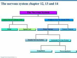
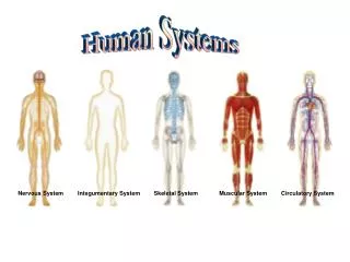
II. Organization of the Nervous System A. Structural Classification- Two Subdivisions: 1. Central Nervous System (CNS) - occupies the dorsal cavity, integration and commanding centers a. includes the brain and spinal cord 2. Peripheral Nervous System (PNS) - contains mainly the nerves that extend from the brain and spinal cord a. includes cranial and spinal nerves
B. Functional Classification- concerned only with the PNS and has two subdivisions 1. Sensory or Afferent division- Nerve fibers that carry information to the central nervous system 2. Motor or Efferent division- Nerve fibers that carryimpulses awayfrom the central nervous system. The Two subdivisions are the somatic and autonomic nervous system. a. Somatic nervous system = voluntary b. Autonomic nervous system = involuntary further divided into the sympathetic and parasympathetic divisions. i. Sympathetic division- the “fight- or-flight” system. Its activity is evident when we are frightened, in emergency, or threateningsituations. ii. Parasympathetic division- sometimes called the “resting and digesting” system. It is most active when the body is at rest and not threatened.
Organization of the Nervous System Sensory neurons Motor neurons
A. Supporting Cells 1. Astrocytes- abundant star-shaped cells, brace neurons, form the boundary between capillaries and neurons, and control the chemical environment of the brain 2. Microglia- spider-like phagocytes that dispose of debris
ALS or Lou Gehrig’s disease, is when nerve cells (neurons) waste away or die, and can no longer send messages to muscles • New research indicates that mutant genes activate microglial cells • the activated microglial cells release proteins that produce nitric oxide or others (cytokines) that cause inflammation, • damage to motor neurons • Accelerates the progress of ALS.
III. Nervous Tissue: Structure and Function A. Supporting Cells 3. Ependymal cells- line cavities of brain and spinal cord, help circulate cerebrospinal fluid
4.Oligodendrocytes- produce myelin sheath around nerve fibers in CNS 5. Satellite cells- protect neuron cellbodies 6. Schwann cells- form myelin sheath in PNS If myelin sheaths are broken down a person develops MULTIPLE SCLEROSIS, More common In women, often diagnosed In the 20’s to 40’s
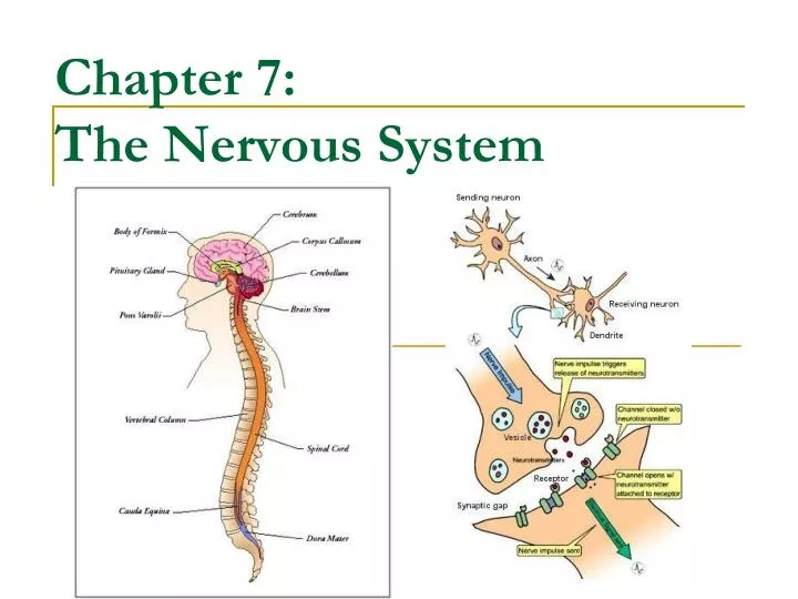
B. Neurons- excitable little cells that make use of their potential! 1. Cells specialized to transmit messages 2. Major regions of neurons: a. Cell body- nucleus and metabolic center b. Processes- fibers that extend from the body 1) dendrites- convey messages towards the cell body 2) axons- conduct messages away from the cell body, axons usually end with terminals that contain vesicles (small sacs) that contain neurotransmitters c. Synapse- the space between two neurons
3. Nerve Fiber Coverings: a. Schwann Cells- produce myelin sheaths- wrapped membranes that enclose the axon b. Nodes of Ranvier- gaps in myelin sheaths along the axon- allows the impulse to travel quickly down the neuron
B. Neurons- excitable little cells that make use of their potential! 4. Functional Classification of neurons- grouped according to the direction the impulse is traveling relative to the CNS a. Sensory or Afferent neurons- carry impulses from sensory receptors. Examples- cutaneous nerve receptors b. Motor or Efferent Neurons- carry impulses from the CNS c. Interneuron (association neurons) - connect sensory and motor neurons Reflex Arc Animation
B. Neurons- excitable little cells that make use of their potential! 5. Structural neuron classification: (determined by the number of processes extending from the cell body) a. Unipolar-have a short process leaving the cell body;found in sensory neurons of PNS b. Bipolar-one axon and one dendrite; found in sense organs (eye, ear) c. Multipolar- many extensions from the cell body; most common type
B. Neurons- excitable little cells that make use of their potential! C. Functional Properties of Neurons 1. Irritability- neurons have the ability to respond to a stimulus 2. Conductivity- the ability to transmit an impulse 3. The plasma membrane at rest is polarized, this is called the Resting potential (-70 mV); this means fewerpositive ions are inside the cell (K+) than outside (Na+). As long as the inside remains more negative than the outside the neuron remains inactive. Resting Membrane PotentialAnimation
Action potential animation • http://www.youtube.com/watch?v=YP_P6bYvEjE • Resting potential • http://highered.mcgraw-hill.com/sites/0072495855/student_view0/chapter14/animation__the_nerve_impulse.html • http://www.blackwellpublishing.com/matthews/channel.html
D. Action Potentials or Nerve Impulses- an electrical charge traveling down a neuron • A nerve impulse begins with a stimulus- usually this is a neurotransmitter released by other neurons, pain receptors, light excites receptors in the eye, etc. • Once the neuron is stimulated the “sodium gates” of the neuron open and sodium ions begin flowing across the cellmembrane. This is called Depolarization: a decrease in membrane potential (inside less negative) increases the chances of an impulse (action potential). • If the action potential starts, it continues down the entire neuron. Action potentials are an “all or nothing” response. • Potassium ions rush out of the neuron after sodium ions rush in, which repolarizes the membrane (returns to resting). A neuron cannot conduct another impulse until repolarization occurs. Action Potential Animation
D. Action Potentials or Nerve Impulses- an electrical charge traveling down a neuron • The propagation of a nerve impulse occurs more rapidly in cells that contain a myelin sheath. This helps the impulse jump from node to node along the length of a neuron. This is called saltatory conduction. Action Potential Animation
Conductivity- How does the impulse travel to the next neuron? • Once the impulse reaches the axon endings, vesicles containing neurotransmitters fuse with the plasma membrane, which ruptures releasing the neurotransmitter (a chemical messenger). • Some neurotransmitters fit receptor sites, some return through reuptake sites and are reused, and some are destroyed by enzymes. • If enough neurotransmitter is released the action potential will continue in the next neuron.
Steps in an ACTION POTENTIAL • Watch the video animation and then use your notes to write down the events taking place http://www.youtube.com/watch?v=oR25iCZ-Eok
Conductivity- How does the impulse travel to the next neuron? F. Reflexes are a predictable, rapid, involuntary response to a stimulus. These responses do not carry the impulse to the brain, they take a shorter path to allow for quicker response. • Reflex Arcs- the direct route from a sensory neuron, to interneuron, to an effector. • Autonomic reflexes include regulation of: smooth muscle, blood pressure, digestive system, and glands. • Somatic reflexes involve the activation of skeletal muscles.
All reflex arcs have five key elements: • sensory receptor- reacts to a stimulus • afferent neurons- carry the message to theintegration center • integration center- the connection between the afferent and efferent pathways • efferent neurons- carry the message away from the integration center • effector organ- gland or muscle stimulated
IV. Developmental Aspects of the Nervous System A. The nervous system begins to form in the first month of embryonic development. Therefore maternal and environmental factors may impair brain development. Mothers that smoke impair the bodies ability to carry oxygen sufficiently which increased the chances of oxygen deprivation to the babies brain cells that are forming. Other severe congenital brain disorders include cerebral palsy which is thought to be caused by a temporary lack of oxygen during delivery. Furthermore, premature babies have trouble regulating their temperature because the hypothalamus is one of the last brain areas to mature prenatally. B. The development of motor control indicates the progressive myelination and maturation of a child’s nervous system. Cerebral refers to the affected area of the brain, the cerebrum, and palsy refers to disorder of movement. Cerebral palsy is caused by damage to the motor control centers of the young developing brain and can occur during pregnancy, during childbirth, or after birth up to about age three.
IV. Developmental Aspects of the Nervous System C. Neurons continue to die throughout life and cannot be replaced. However, most healthy aged people maintain optimal intellectual functioning. Cardiovascular disease is the major cause of declining mental function with age.