Download
1 / 7
70 likes | 193 Views
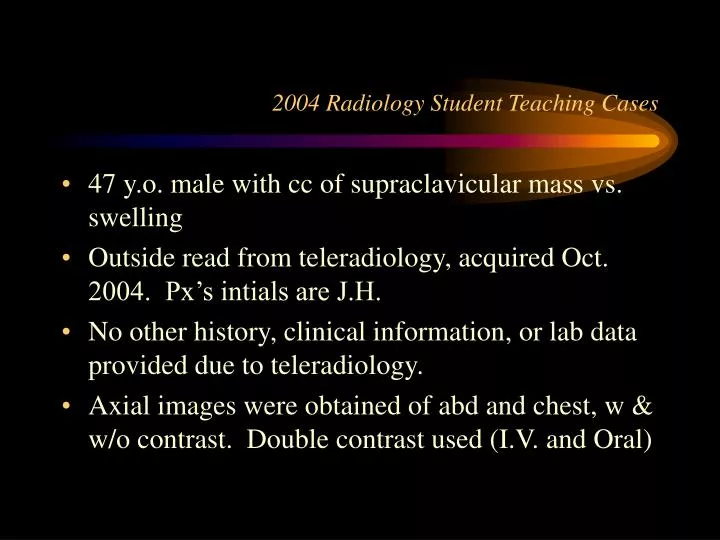
2004 Radiology Student Teaching Cases. 47 y.o. male with cc of supraclavicular mass vs. swelling Outside read from teleradiology, acquired Oct. 2004. Px’s intials are J.H. No other history, clinical information, or lab data provided due to teleradiology.

E N D
2004 Radiology Student Teaching Cases • 47 y.o. male with cc of supraclavicular mass vs. swelling • Outside read from teleradiology, acquired Oct. 2004. Px’s intials are J.H. • No other history, clinical information, or lab data provided due to teleradiology. • Axial images were obtained of abd and chest, w & w/o contrast. Double contrast used (I.V. and Oral)
Non-contrast axial image of the thorax, bone window. Note the calcifications in the mediastinum.
Contrast axial image of the thorax, bone window. Internal mammary system Collateralized flow (internal mammary and subcostal) Contrast in the rapidly narrowing SVC Azygos vein Note the presence of contrast in the collateralized vessels before the appearance of contrast in the descending aorta.
Post-Contrast Axial Image Almost completely occluded SVC
Fibrosing Mediastinitis • extensive fibrosis following granulomatous infections of mediastinal nodes. • Common sequelae include SVC syndrome and obstuction of the pulmonary vasculature. Lymphadenopathy may be clinically evident. • Two main pathophysiologies: • Post-infectious (usually TB or Histoplasmosis) • Idiopathic and associated with retroperitoneal fibrosis • Associated radiologic findings include: calcification of the mediastinal and hilar lymph nodes and collateralization of venous system. (Grainger & Allison’s Diagnostic Radiology. 4th Ed. Pg. 372)
In terms of radiologic workup, CT is the best modality, because MRI cannot reliably detect calcifications. VQ scans can be helpful in determining the extent of pulmonary compromise. • The prognosis of fibrosing mediastinitis is poor. (Cummings: Otolaryngology: Head and Neck Surgery, 3rd ed. Pg 2361) • Many patients demonstrate nonprogressive disease without major disability, and do well without therapy.(Cohen & Powderly: Infectious Diseases, 2nd ed. Pg. 434) • Treatment: treat underlying disease (i.e.-itraconazole for Histo, standard multi-agent regimen for TB), surgical decompression if indicated.
Final Verdict? Upon CT examination of the abdomen, prominent adenopathy was noted in the retroperitoneal space. In the setting of a lack of clinical information concerning previous infectious granulomatous disease, the presumed diagnosis is therefore idiopathic retroperitoneal fibrosis with associated fibrosing mediastinitis.