Download
1 / 64
640 likes | 771 Views
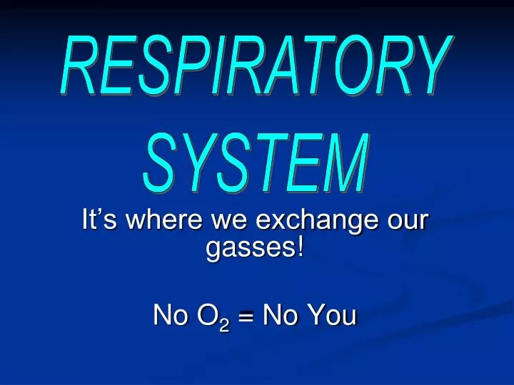
RESPIRATORY SYSTEM. It’s where we exchange our gasses! No O 2 = No You. Respiratory System Functions. Homeostatic mechanism Air Distributor Gas exchanger Filters, Warms, & Humidifies air Sinuses Influence speech/sound Make smell possible. Respiratory Structure.

E N D
RESPIRATORY SYSTEM It’s where we exchange our gasses! No O2 = No You
Respiratory System Functions • Homeostatic mechanism • Air Distributor • Gas exchanger • Filters, Warms, & Humidifies air • Sinuses • Influence speech/sound • Make smell possible
Respiratory Structure • It’s designed as a tube with many branches ending where gasses are exchanged • Gas exchange works by diffusion • Continually smaller branches gets air close enough to the blood to diffuse
Respiratory Tracts • Upper Respiratory Tract • Nose, Pharnyx, & Larynx • Lower Respiratory Tract • Trachea, all segments of Bronchial Tree, & lungs
Respiratory Mucosa • Membrane that lines most of the air distribution tubes • Serves as an air purification system • Removes contaminants from air • ie: insects, dust, pollen, bacteria • 125 ml + of mucus is produced daily • A “mucus blanket” covers the lining of air distribution tubes
Respiratory Mucosa • Cleansing mucus moves upward from the bronchioles toward the pharnyx on cilia • Cilia move in only one direction • Cigarette smoke paralyzes the cilia • Results in the smoker’s cough!
Upper Respiratory Tract
Nose • Where air enters the r.t. • Flows into L & R nasal cavities • Nasal septum separates • Paranasal sinuses drain into the cavities • Frontal, maxillary, sphenoidal, & ethmoidal • Hollow spaces help lighten skull bones • Serve as resonant chambers for sound production • Lined with mucus membranes
Lacrimal Sacs Drain into nasal cavity from tears Conchae ↑ surface area over which air must flow Warms & humidifies air Nose
Pharnyx • Commonly called the throat • Approx. 12.5 cm long • Consists of 3 parts • Nasopharnyx • Oropharnyx • Laryngopharnyx • Is a passage for food & air on its way to the stomach & lungs
Pharynx • Tonsils • Lymphatic tissue embedded in pharnyx • Pharyngeal Tonsils (Adenoids) • Located in nasopharynx • Palatine Tonsils • Located in oropharnyx • Tonsillitis – inflammation of tonsils • Treated with antibiotics • Removed only as last resort
Larynx • Commonly called the “voice box” • Composed of several pieces of cartilage • Largest of these is the “adam’s apple” • Vocal Cords • 2 short fibrous bands stretched across the interior of the larynx • Muscles control the tenseness of the bands • This changes the pitch as air passes over it
Larnyx • Glottis • Space between vocal cords • Epiglottis • Partially covers the opening of the larynx • Acts as a trapdoor • Closes larynx during swallowing preventing food from entering the trachea
Upper Respiratory Tract Disorders • Infections in the nose, pharnyx, & larnyx • Upper Respiratory Infection (URI) • Rhinitis (Coryza) • Inflammation of the nasal mucosa • Pharyngitis (Sore Throat) • Inflammation of the pharnyx • Strep Throat
Upper Respiratory Tract Disorders • Laryngitis • Inflammation of the mucous lining in the larynx • Swelling of vocal cords
URT Anatomical Disorders • Deviated Septum • Nasal septum strays from the midline of the nasal cavity • Epistaxis • Nose bleeds
Lower Respiratory Tract
Trachea • Commonly called the “Windpipe” • About 11 cm long • Goes from larnyx to the bronchi • Passageway for air to reach the lungs • Has 15 – 20 C-shaped rings of cartilage for strength • Choking occurs when the trachea is obstructed
Bronchi • The 1st branches from the trachea leading to the lungs • Primary Bronchi • In each lung, the primary bronchi branch into smaller & smaller tubes • Secondary bronchi
Bronchioles • Tiny tubes whose walls contain only smooth muscle
Alveolar Ducts • Microscopic tubes which resemble the main stem of a bunch of grapes
Alveolar Sacs • The end of each alveolar duct has this • Resembles a cluster of grapes
Alveoli • Makes up the wall of each alveolar sac • Resembles a single grape • Very effective in gas exchange • Extremely thin walled • Each lies in contact with a capillary • Millions contained in each lung
Surfactant • A substance covering the inside of the alveolus • Reduces surface tension • Keeps alveoli from collapsing
Respiratory Distress
Infant Respiratory Distress Syndrome (IRDS) • Affects prematurely born infants of less than 37 weeks gestation or weigh less than 5 lbs at birth • Caused by Lack of surfactant in the lungs
Adult Respiratory Distress Syndrome (ARDS) • Caused by impairment or removal of surfactant in the alveoli • Accidental inhalation of foreign substances • Water, vomit, smoke, chemical fumes
Pleura • Pleura covers the outer surface of the lungs & lines the inner surface of the rib cage • Parietal pleura • Lines the walls of the thoracic cavity • Visceral Pleura • Covers the lungs • Intrapleural space • Lies between the two pleural membranes
Pleurisy • Inflammation of the parietal pleura • Characterized by difficulty in breathing & stabbing pain • There is discomfort & restriction in breathing • Caused by rubbing between the two pleura
Atelectasis • Collapse of the lung for any reason • While collapsed, the lung is useless in breathing • Pneumothorax • Puncture wound to the chest wall or rupture or the visceral pleura • Literally means “air in the thorax” • Hemothorax • Presence of blood in the pleural space
Respiration • Exchange of gases (o2 & CO2) between a living organism and its environment • External (pulmonary) respiration • Process that moves air in & out of the lungs • Internal Respiration • Exchange of gases between the blood & cells of the body
Inspiration • Moving air into the lungs • Diaphram – primary muscle • Contracts - ↑ volume - ↓ air pressure – causes air to rush into lungs
Expiration • Moves air out of the lungs • Diaphram • Relaxes - ↓ volume – ↑ air pressure – forces air out of the lungs
How is it Measured? • Spirometer • A special device you breathe into to measure the amount of air exchanged in breathing
Types of Volumes • Tidal Volume (TV) • The amount of air we regularly breathe in & out • Approx. 500 mL (about a pint) • Vital Capacity (VC) • The largest amount of air that we can breathe out in one expiration
Types of Volume • Expiratory Reserve Volume (ERV) • The amount of air that can be forcibly exhaled after expiring the TV • Inspiratory Reserve Volume (IRV) • The amount of air that can be forcibly inspired over & above a normal inspiration • VC = TV + IRV + ERV
Types of Volumes • Residual Volume (RV) • The air that remains in the lungs after the most forceful expiration