Download
1 / 18
180 likes | 317 Views
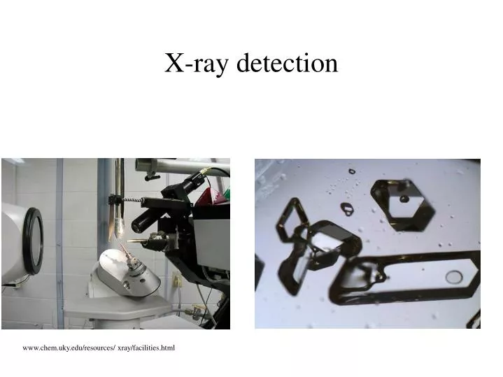
X-ray detection. www.chem.uky.edu/resources/ xray/facilities.html. How can we solve structures. X – ray detection. Normally the best. NMR. Not very precise. Looks like > 3 Å X-ray structures. Only small proteins (80 AA).

E N D
X-ray detection www.chem.uky.edu/resources/ xray/facilities.html
How can we solve structures • X – ray detection. Normally the best. • NMR. Not very precise. Looks like > 3 Å X-ray structures. Only small proteins (80 AA). • Electron microscopy (> 7 Å) not useful for models and only the shape is seen, no details. • Other spectroscopic methods sometimes are useful for indirect information, or very local information. E.g. EXAFS can give very detailed information about metal contacts.
Problems with PDB files • The watermolecules often are just guessed, because they are difficult to see. • C, N, and O cannot be distinguished. • Hard to distinguish ion from water from alternate structure of amino acid side chain. • Resolution > 1 Å • Difficult on big proteins > 200 AA
Discussion • Advantage: • Better than everything else • Because of the water molecules in the crystal there is a good congruence to the structure in the water surrounding • Disadvantage: • Difficulties in fabrication the crystals, specially membrane crystals are very difficulty to make.
PDB • Protein Data Bank (PDB) • URL: http://www.rcsb.org/pdb/ • All coordinates of the proteins are collected there • Files are structured and formatted so easy to search • Contains all structures from X-ray and NMR • Older than the most databanks • Because it is historic it is very unflexible
HEADER TRANSFERASE (METHYLTRANSFERASE) 05-JAN-96 1VID TITLE CATECHOL O-METHYLTRANSFERASE HELIX 1 1 LYS 5 ASN 16 1 12 HELIX 2 2 PRO 22 GLN 35 1 14 HELIX 3 3 GLY 43 TYR 57 1 15 HELIX 4 4 TYR 71 LEU 79 1 9 HELIX 5 5 PRO 93 ALA 106 1 14 HELIX 6 6 SER 119 TYR 130 1 12 HELIX 7 7 LYS 144 LYS 156 5 13 HELIX 8 8 PRO 177 GLY 185 1 9 SHEET 1 A 7 PHE 189 TYR 197 0 SHEET 2 A 7 VAL 204 TYR 212 -1 N ILE 211 O GLU 190 SHEET 3 A 7 VAL 165 ALA 168 -1 N ALA 168 O GLU 208 SHEET 4 A 7 MET 137 LEU 140 1 N VAL 138 O VAL 165 SHEET 5 A 7 LEU 61 LEU 65 1 N LEU 63 O MET 137 SHEET 6 A 7 ARG 85 GLU 90 1 N ARG 85 O VAL 62 SHEET 7 A 7 VAL 112 ASN 116 1 N THR 113 O LEU 86 ATOM 1 N THR 4 -32.796 52.984 34.270 1.00 25.07 N ATOM 2 CA THR 4 -32.168 53.250 35.602 1.00 25.39 C ATOM 3 C THR 4 -32.658 54.592 36.116 1.00 24.95 C ATOM 4 O THR 4 -33.177 55.399 35.337 1.00 25.40 O ATOM 5 CB THR 4 -30.631 53.322 35.498 1.00 26.05 C ATOM 6 OG1 THR 4 -30.256 54.471 34.725 1.00 26.33 O ATOM 7 CG2 THR 4 -30.077 52.059 34.847 1.00 26.84 C PDB Format
B - factor • The PDB databank gives also the B-factor • Important for modelling • Gives information about how precise the residue is located. B >= 100 means the atom exists but cannot be seen; B >=60 means atomic position is doubtful
ATOM 2 CA THR 1 16.967 12.784 4.338 1.00 10.80 ATOM 3 C THR 1 15.685 12.755 5.133 1.00 9.19 ATOM 4 O THR 1 15.268 13.825 5.594 1.00 9.85 ATOM 5 CB THR 1 18.170 12.703 5.337 1.00 13.02 ATOM 6 OG1 THR 1 19.334 12.829 4.463 1.00 15.06 B-factor • Last value in the row • B-factor • a measurement for the atomic position • measured in Å2. 80 Å2 is about a RMS of 1 Å.
Example • 1crn • ATOM 203 C TYR 29 1.125 11.125 7.815 1.00 4.92 1CRN 272 • ATOM 204 O TYR 29 .286 10.632 8.545 1.00 7.13 1CRN 273 • ATOM 205 CB TYR 29 .755 11.229 5.322 1.00 9.66 1CRN 274 • ATOM 206 CG TYR 29 -.203 10.044 5.354 1.00 11.56 1CRN 275 • ATOM 207 CD1 TYR 29 -1.547 10.337 5.645 1.00 12.85 1CRN 276 • ATOM 208 CD2 TYR 29 .193 8.750 5.100 1.00 14.44 1CRN 277 • ATOM 209 CE1 TYR 29 -2.496 9.329 5.673 1.00 16.61 1CRN 278 • ATOM 210 CE2 TYR 29 -.801 7.705 5.156 1.00 17.11 1CRN 279 • ATOM 211 CZ TYR 29 -2.079 8.031 5.430 1.00 19.99 1CRN 280 • ATOM 212 OH TYR 29 -3.097 7.057 5.458 1.00 28.98 1CRN 281 • ATOM 213 N THR 30 2.470 10.984 7.995 1.00 5.31 1CRN 282 • ATOM 214 CA THR 30 2.986 9.994 8.950 1.00 5.70 1CRN 283 Poor alignment caused by Bill Gates who doesn’t understand the Courier font.
Examples • 1ycq, 2,3 Ångstrom resolution, very bad • 1chc: no resolution, very suspicious • 1rdb, 1,9 Angstroem, better but B-values are high • 2frt, is a model • 1frt, 4,5 Angstroem resolution, no discussion