Download
1 / 31
330 likes | 793 Views
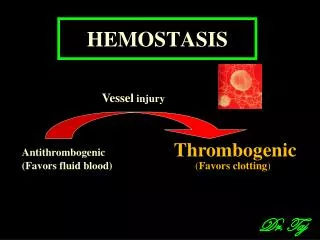
Hemostasis system. Hemostasis system. Hemostasis is the physiologic system, which support the blood in the fluid condition and prevent bloodless. Hemostasis system vital necessary and functionally connect with the cardiovascular, digestive, breathing, endocrine and other systems.

E N D
Hemostasis system • Hemostasis isthe physiologic system, which support the blood in the fluid condition and prevent bloodless. • Hemostasis system vital necessary and functionally connect with the cardiovascular, digestive, breathing, endocrine and other systems.
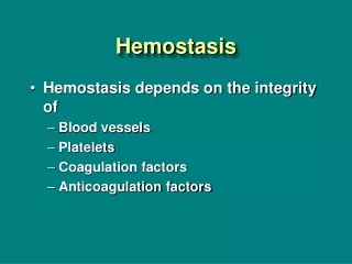
The components of hemostasis system • The components of hemostasis are • - wall of the vessels, • - blood cells – platelets, erythrocytes, leukocytes, • - enzymes and nonenzymes components of plasma – clotting and anticlotting substances, fibrinolytic components. • There are 2 kinds of hemostasis: vessel-platelets (primary) and coagulative (secondary).
Platelets • Properties and function of platelets • Quantity of platelets is 180-320 G/L. • Diameter of platelets is 1-4 micrometers, thickness – 0,5-0,75 micrometers. • They are the little peace of megakaryocytes cytoplasm (from one megacariocytes may develop few hundred of platelets). Platelets circulates in blood from 5 to 11 days and than destroyed in liver, lungs, spleen by the cells of macrophages system. Formation is regulated by thrombopoietin. • Their granules contain serotonin, Ca2+, enzymes, ADP, and platelet-derived growth factor (PDGF) • Platelets function in the clotting mechanism by forming a temporary plug that helps seal breaks in blood vessels • Platelets not involved in clotting are kept inactive by Nitric Oxide (NO) and prostaglandins.
Function of platelets are: • 1. haemostatic function – platelets produce substances, which are secure the hemostasis. • 2. Angiotrophic function – provide trophy of endotheliocytes of vessel wall, support structure and functions of micro vessels. These function is realize by adhesion of platelets to endotheliocytes and injection the enzymes into the endotheliocytes. For one day near 35 G/l of platelets do this function. • 3. Transport function – transfer the enzymes, ADP, serotonin and other. • 4. Phagocytes function – the contain of platelets help to kill viruses and antigens bodies. • 5. Regeneratory function – platelets have the growth factor, which help to grow the endothelial and muscles cells which are present in the vessel wall.
Stages of vessel-platelets hemostasis • 1. Shorting spasm of the vessels – vascular spasm duration to 1 minute is caused by catecholamines and other enzymes. Diameter of vessels decrease on ½-⅓. Mechanism of it development determine by secretion of serotonin and thromboxan A2 from platelets and epinephrine from ending of sympathetic nerves. • 2. Adhesion of platelets – activation of platelets and stick it to the place of defect in vessel wall. • 3. Reverse aggregation of platelets – the thrombus which are formed may make way for plasma. • 4. Unreverse aggregation of platelets – the thrombus which are formed can not may make way for plasma. • 5. Retraction of platelets plug – decrease the size of plug, pack down the plug.
Investigation of vessel-platelets hemostasis • 1. Calculation of the platelets quantity 180-320 G/l. • 2. Determination of duration of capillary bleeding after Duke’s method – to 3 minute in norm. • 3. Sample of fragility of capillary – to 10 petechias in norm in a round with diameter 5 centimeters.
Coagulative (secondary) hemostasis. • Characteristics of clotting factors • There are 12 clotting factors: • I – fibrinogen; • II – prothrombine; • III – thromboplastin of tissue; • IV – ions of calcium; • V – proaccelerin; • VII – proconvertin; • VIII – antihemophylic factor A; • IX – Christmas factor or antihemofilic factor B; • X – Stuart-Prower factor or prothrombinase; • XI – plasma thromboplastin antecedent; • XII – Hageman factor; • XIII – fibrin stabilizing factor.
Some of them are enzymes – II, VII, IX, X, XI, XII,XIII; otherare not – I, III, IV, V, VIII. The vitamin K is necessary for the functional activity of II, VII, IX, X factors. • Vitamin K (produced by bacteria in the colon) stimulates the production of prothrombin by the liver.
Regulation of the clotting mechanisms • Increase of clotting names hypercoagulation, decrease – hypocoagulation. Hypercoagulation may be in a stress cases. It depends on epinephrine, which concentration increased in the cases of stress. Epinephrine increase from the vessels walls factors from which produced prothrombinase. In cases of big concentration epinephrine should activate XII factor in a bloodstream. It divides fats and fat acids, which have prothrombinase activity. After the hypercoagulation stage may be secondary hypocoagulation.
Coagulogram -Time of clotting by Ly-Wait – 5-10 minutes; - Time of plasma recalcification – 60-120 seconds; - Thrombotest – IV, V, VI degree; • Thromboplastin time – 12-15 seconds; • Thromboplastin index – 80-105 %; • Concentration of fibrinogen – 2-4 g/L; • Tolerance of plasma to heparin – 6-11 minutes; • Heparin time – 50-60 seconds; • Fibrinolysis – 15-20 %.
Anticoagulative mechanisms. Fibrinolysis. • The primary anticoagulants are • antithrombin III (the most important anticoagulant in the blood); • heparin (originally found inthe liver, by large basophilic cells in tissues of various organs. Heparin reduces the ability of the blood to clot by blocking the changeof prothrombin to thrombin. It can also be used to aid in reducing clots in cases in which internal clotting has already occurred. Heparinform complex with antithrombin-III. Activate nonenzyme fibrinolysis.);
alpha-2-macroglobulin (Alpha-2-macroglobulin is a similar to antithrombin-heparin cofactor in that it combines with the proteolytic coagulation factors. Its activity is not accelerated by heparin. Its function is mainly to act as a binding agent for the coagulation factors and prevent their proteolytic action until they can be destroyed in various ways. It a faint inhibitor of thrombin, connect with plasmin), • alpha-1-antitripsin (Alpha-1-antitripsin inhibits thrombin activity, IXa, XIa, XIIa factors, plasmin and kallilrein), • protein C (Protein C inhibits VIIIa, Va factors. It activity depend of thrombin and vitamin K concentration).
Functionation of secondary anticlotting substances • Primary anticoagulants are produce and present all time in plasma and secondary anticoagulants form in a case of blood clotting. They are antithrombin-I or fibrin and products of fibrinolysis or products of fibrinogen degradation. Fibrin is sorbs and inactivates thrombin and Xa factor. Products of fibrinolysis inactivate ending stage of clotting, IXa factor, platelets' aggregation.
Fibrinolysis • Clot dissolved by activity of plasmin, an enzyme which hydrolyzes fibrin