Download
1 / 44
500 likes | 552 Views
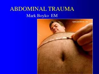
ABDOMINAL TRAUMA. ABDOMINAL VISCUS. Erect position exposes the abdomen More effective with relaxed muscles Some organs protected by bony structures ) Some organs project in thorax or pelvis and can be injured in trauma of these regions

E N D
ABDOMINAL VISCUS • Erect position exposes the abdomen • More effective with relaxed muscles • Some organs protected by bony structures) • Some organs project in thorax or pelvis and can be injured in trauma of these regions - Any trauma bellow angle of the scapula (post) or bellow the niple anterior can injure abdominal organs
CONCUSSION • Causes • Road accidents – 2/3 • Work accidents • Agresion • Sport • Varia
CONCUSSION • ♂ young • Very frequent • Politrauma • Rural accidents
concusion Classification according to type of injury • Blast injury • Mixt mechanism • Direct trauma - simple - crush • Indirect trauma - counter hit
concussion • Type of agent • Solid • Explosive waves • Bone fragments • Significant factors • Agent • Speed • Force • Direction: perpendicular/tangential • Surface • Natural visceral protection • Associated diseases
concussion Clasification: pathology • Parietal lesions • Morel-Lavalle - supraaponeurotic hematoma • Muscle ruptures – hematoma→ properitoneal hematoma - posttraumatic hernia • Associated bone lesions
Seroma Morell Lavalle • Small vessels injury with spontaneous hemostasis – tangential trauma with shearing mechanism • Develops in time, but does not feel the entire space available = fluctuence not always present • Usually normal skin • Will be absorbed in time, sometimes requiring aspiration
Hematoma – rectus abdomini rupture • Anatomic particularities: • Fascial intersections that segment the muscle • Rectus sheet • Abundant network of vessels , large vessels inside the sheet • Hematoma is well circumscribed, in tension, developed between two intersections. • During contraction of the wall: painful and does not disappear inside the abdomen. • Diagnostic: sudden onset, related to trauma are fundamentals in understanding the diagnosis.
Hematoma –psoas muscle rupture • Anatomic particularities: • Situated deep in retroperitneum • Adjacent to branches from the lumber plexus) • Developed in the retroperitoneum • Disappears during abdominal wall contractions • Diagnostic: sudden onset, related to trauma are fundamentals in understanding the diagnosis • May appear spontaneous
Rupture of the diaphragm • Indirect mechanism via an acute increase in abdominal pressure • Direct mechanism – crushing the base of the thorax • False herniation of abdominal viscus in the thorax. (false = no peritoneum) • Respiratory problems due to intrathoracic compression • Digestive problems – difficult to evaluate in a trauma patient with more serious lesions.
Posttraumatic hernia • Early or late complication of trauma • BREAK IN THE MUSCULAR-FASCIAL LAYER – may be obscured by gravity of initial trauma • In time it develops like a true hernia through a new week point • Symptoms are very similar to all postoperative hernia BUT no scar
concussion 2. Intraabdominal organ lesions • Cavitary – 1/3 • Small bowell ruptures – most frequent - ruptures - complet - incomplet - secondary perforations - posttraumatic stenosis • Stomach – more often on a full stomach
concussion • Duodenum – DII, DIII - Retroperitoneal spillage: bile, blood and gas • Colon – - peritoneum - retroperitoneum VERY SERIOS: FECAL PERITONITIS
concussion • Bile ducts- gallbladder - CBD COLEPERITONEUM • Bladder– 3% (intra or extraperitoneal)
concussion B. Parenchimatous organs • Fissure, ruptures, avulsion of pedicle • Hematoma: - subcapsular - central • Liver – 25% • Spline – 50% • Pancreas – 5% • Kidney – 10% →! Hemorhagein 2 seq
Superficial Penetrate Perforated 20% of all peace time abdominal trauma 90% of all war time abdominal trauma Classification Wounds and contusions can be present in the same time
Non-penetrated abdominal wounds • Diagnostic is essential = lack of penetration • Intact serosal layer – difficult to appreciate especially in a blunt trauma with a wound
DIAGNOSTIC CRITERIA • Anamnesis • Weapon and trajectory • Relative position of aggressor and victim • Direction of the weapon as it hits • Physiologic status • Number of wounds
Local examination • Gentle, after a careful antiseptic preparation of the skin and wound • Use a blunt gentle instrument to probe the wound • If not a simple stab wound (that is complex wounds with non-linear trajectory) the information will always be incomplete. ATTENTION to strata movements between impact and examination • An examination with a negative result is not necessary conclusive
General examination of the abdomen • Look for signs and symptoms suggestive for penetrating and perforated wound • Monitor the clinical status of the patient – that is safe in the case of a negative evaluation (regarding a probable superficial wound) • Admit patient for hospital care for at least 24 hours
Lab exams • Their purpose is to identify signs of major syndromes related to the peritoneal cavity • Peritonitis • Hemorrhage • Intestinal obstruction • Acute pancreatitis • According to type of wound and trajectory
Surgical evaluation of the abdominal cavity • In this case IT IS a method of EXPLORATION • Laparotomy • Laparoscopy MAJOR LIMITS • Check the integrity of the peritoneal surface • Check the integrity of viscus • Check for fluid in periteneum TYPE • Andominal exploration should be as complete as possible – HOW MUCH IS COMPLETE
TAKE GOOD CARE • Any abdominal wound (even very small or apparently without significance) can be penetrated. MINIMAL ACCESS SURGERY • A small wound can be accompanied by a big disaster in the abdomen. • Initial evaluation can be misleading
Penetrated wounds • All the abdominal wall has been penetrated (including the parietal peritoneum) but no viscus in injured • It is not common – more frequent with stab wounds • Exploration – same methods
Clinical evaluation • Wound exploration: • How much the instuments can be inserted in comparison with the width of the abdominal wall = RELATIVE • Is the probe free to move? = RELATIVE • If the wound is large enough abdomina viscus can herniate outside = DIAGNOSTIC
Significance • Major risk for a viscus injury, even if not apparent • Major risk to err due to absence of clinical manifestation at presentation • Risk of infection of the peritoneal cavity • In traumatic evisceration – risk of strangulation
Perforated wounds • Symptoms depend on viscus involved and time interval from lesion (25-35% multiple organs affected) • Dg obvious when in the wound • Digestive content OR colonic content OR blood in quantity larger then we expect ????? • Symptoms develop in time – check for patients condition
MAJOR LESIONS: cavitary organs 1. Stomach • Concussions: simple hematoma – to dilaceretion • Gastric wounds: anterior or posterior wounds
MAJOR LESIONS: cavitary organs 2. Duodenum - simple concussions - intramuralhematoma: may develop instestinal obstruction, perforation, or nothing - rupture: complete or incomplete; total or partial • intraperitoneal (cu peritonită) • retroperitoneal • - duodenal wounds
MAJOR LESIONS: cavitary organs 3. Small bowel and mesentery – most frequent - Hematoma of the intestinal wall: may develop obstrution, perforation or resolution. PERFORATION IS IN 2 SEQUENCES - ruptures • wounds – a wide range of complexity • Hematoma and ruptures of mesentery: may affect the bowell and may produce massive bleeding
MAJOR LESIONS: cavitary organs 4. Colonuland mesocolon: same as small bowell but content more septic 5. Rectum • - penetrating wounds: trauma of the pelvis, gunshot, falling in sharp objects • - iatrogenic trauma • - unusual causes ingested foreign body foreign bodies introduced in the rectum • - explosions following hyperinflation (strange jokes, psyhopatic behavior).
MAJOR LESIONS: solid organs 1. Liver a) Primary lesions of the parenchim: • - Subcapsular hematoma – preservation of the capsule which can retain large volumes: major risk for secondary rupture (hours – days) secondary hemoperitoneum • - Wounds and ruptures ; • -Avulsion b) Lesions of the hepatic pedicle: gallbladder, CBD, major vessels
MAJOR LESIONS: solid organs 2. Spline • ruptures and wounds; • Subcapsular hematom – secondary rupture in peritoneal cavity • Avulsion of the pedicle 3. Pancreas – unusual 1-2% - crash usually - simple concussion -rupture with small duct lesions; - rupturewith Wirsung duct lesion; - crushing Major problem: pancreatitits
MAJOR LESIONS: solid organs D. Retroperitoneal hematoma May associate: • Pelvic fractures or vertebral fractures; • Injury of the bug vesseks in the retroperitoneu • Trauma of the adrenal glands • Trauma of the kidney. E. Kidney trauma a) Renal parenchime • - hematoma with intact capsule; • - fissure with broken capsule; • - dileceration of renal parenchime • b) Renal pedicul: elements of the pedicul including the urinary system,
III. DIAGNOSTIC • Evalution of vital function • Conscience • Full examination • Hierarchy of lesions • Repeated examination: dynamic of lesions
III. DIAGNOSTIC 1.ANAMNESIS
2. Clinical examination • Signs of hemodynamic instability • Abdominal wall lesions • Major abdominal syndromes PLUS: - large bore venous access - naso-gastric aspiration - urinary catheter
CLINICAL EXAMINATION * inspection * palpation * percussion * auscultation * Rectal/Vaginal examination
3. Evaluation a) hematoogcal b) chemistry c) radiology - plain abdominal X-Ray - thoracic X-Ray: must be done in abdominal wounds - water soluble digestive studies - intravenous urography - cystografy: lesions of the bladder - diagnsotic peritoneal lavage: careful to contraindications (obstructions, adhesions)
3. Evaluation a) US b) CT scan (spiral) c) Laparoscopy
TREATMENT • Catastrophe management • Individual care for each involved organ • Hemostasis • Resections • Suturing • etc