Download
1 / 60
600 likes | 606 Views
Dynamic DNA Notes. (Ch 16). Science as a Process:. Many scientists contributed to current knowledge of DNA structure and function. Here are some highlights. A. Morgan (remember fruit flies). Determined that genes are on chromosomes Chromosomes are made of DNA and protein

E N D
Dynamic DNA Notes (Ch 16)
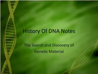
Science as a Process: • Many scientists contributed to current knowledge of DNA structure and function. Here are some highlights.
A. Morgan (remember fruit flies) • Determined that genes are on chromosomes • Chromosomes are made of DNA and protein • Unknown: Is DNA or Protein the genetic material • Best candidate at the time: proteins • *great heterogeneity • *specificity of function • *little known about DNA
B. Griffith (1928) - studied strep pneumoniae bacteria • S strain - living bacteria that is pathogenic (able to cause disease in mouse) • R strain - mutant form, nonpathogenic • Heat Killed S cells - Harmless
B. Griffith (1928) - studied strep pneumoniae bacteria • 1. Inject living S cells into mouse - mouse dies • 2. Inject living R cells into mouse - mouse lives • 3. Inject heat killed S cells into mouse - mouse lives • 4. Mix heat killed S cells with living R cells - mouse dies and living S cells are found in it’s blood
Griffith’s conclusion: • Transformation occurred. Some portion of dead cells was transformed into living cells • Unknown: What is the transforming agent
C. Avery, McCarty, MacLeod (1944) • 1. Purified various chemicals from the heat killed bacteria and attempted to transform the living R cells • Conclusion: Only DNA was able to transform the R cells into pathogenic cells so DNA must be the transforming agent
D. Hershey and Chase (1952) - • Studied bacteriophages (virus that infects a bacteria) - T2 infects the bacteria e. coli • They wanted to confirm that DNA is the transforming agent. Viruses are made of DNA (or RNA) surrounded by a protein coat
1. Tag DNA with a radioactive isotope of some T2 phages with Phosphorous (only found in DNA) • 2. Tag the protein of different T2 phages with radioactive sulfur (only found in protein) • 3. Allow the T2 tagged with phosphorous to infect e. coli
4. Allow the T2 tagged with sulfur to infect other e coli. • 5. Put in blender, centrifuge and force bacteria to bottom of test tube leaving virus parts on top (they are lighter) • 6. Test the bacteria at bottom of tube for radioactivity and the virus parts on top
Conclusion: • Only radioactive phosphorus was found in the bacteria and radioactive sulfur remained with the virus parts on top. • Therefore, DNA was being injected into the bacteria so DNA is the transforming agent.
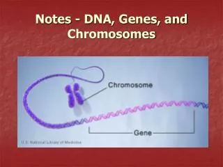
E. More Circumstantial evidence for DNA • 1. DNA is exactly doubled before mitosis • 2. DNA is distributed equally to daughter cells after mitosis • 3. Diploid cells have exactly twice as much DNA as haploid gametes • 4. DNA varies from one species to the next
5. DNA has molecular diversity - there are varying amounts of nitrogen bases (amount of A, G, T and C are not equal) • 6. Chargaff’s rule - the amount of A is about the same as T, amount of G is same as C • This evidence is further proof of DNA as genetic material
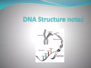
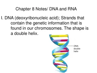
Structure fits Function • A. Watson and Crick (1953) - used as photograph done by X-ray crystallography to help determine the structure of DNA
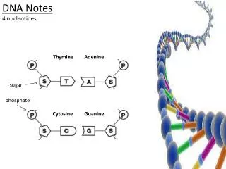
Watson and Crick • *DNA is a Double Helix (like a twisted ladder) composed of 2 strands • *Once they knew the shape, they had to determine how the sugar, phosphate and base fit into the structure to give it the proper shape • *Backbone - sugar-phosphate (sides of ladder) • *Middle - nitrogen bases (rungs of ladder) • *Twist every 10 base pairs
Watson and Crick • *Purines (2 rings) can’t pair together because the double helix would be too wide • *Pyrimidines (1 ring) can’t pair together because the double helix would be too narrow • *Adenine pairs with Thymine making 2 hydrogen bonds • *Cytosine pairs with Guanine making 3 hydrogen bonds (this explains Chargaff’s rule) • *The bases can be in any order along the chain, resulting in the huge variety of DNA found in different species
B. DNA Replication • The structure of DNA (A-T, G-C) also allows for copying mechanism (form fits function)
1. Semiconservative Model for replication • *Each original strand serves as a template for the replication of a complementary strand
Original Strand Separates and becomes template A-T A T G-C G C T-A T A C-G C G C-G C G A-T A T G-C G C 1. Semiconservative Model for replication
2. Conservative Model: • Parent double helix remains intact while a second, all new copy is made
3. Dispersive model: • Each strand of old and new DNA contains a mixture of old/new DNA
*The semiconservative model was proposed by Watson and Crick • *Meselson and Stahl (late ‘50’s) tested the three possible models of replication by labeling the parent DNA with radioactive Nitrogen, allowed replication to occur. • Centrifuged and checked for radioactivity.
Found a hybrid of radioactive and regular DNA. When replicated again, they found both regular DNA and hybrid DNA. • Conclusion: DNA replicates by semi-conservative replication
C. Methods of Replication • Very precise process (one error per billion nucleotides) • Occurs in a short period of time. • Process involves many enzymes and other proteins
1. Origin of Replication • site where DNA begins replicating • *bacteria - have only one origin of replication • *eukaryotes - hundreds/thousands of origins of replication • *Form a bubble and replication occurs in both directions using the parent strands as templates. At end of bubble is a replication fork
2. DNA polymerases • enzymes that catalyze the elongation of DNA by adding the correct base to match the template (50-500 per second)
3. Energy • substrate is a nucleoside triphosphate with 3, high energy phosphate bonds attached to the nitrogen base • *As the base gets added, it loses two phosphate groups that provide the energy needed to form DNA. • The other phosphate and the sugar become the backbone of the ladder
4. Strands are Antiparallel • run in opposite directions (see picture) • *DNA polymerase can only add nucleotides to a 3’ end, not to the 5’ end so DNA can only grow in the 5’3’ direction. This side of the DNA is called the Leading Strand
*The other strand grows away from the replication fork and is called the lagging strand because it can only add new nucleotides in small segments, as the bubble grows • *The lagging strand is created in small fragments called Okazaki Fragments. These must be joined together by an enzyme called DNA Ligase
5. DNA Polymerase can only add to an existing nucleotide • (it can’t start synthesis on it’s own). • *To solve this problem, a short stretch of RNA is added at the origin of replication called a Primer. An enzyme called Primase joins the RNA bases together.
*For the leading strand, only one primer is needed because the DNA grows continuously • *For the lagging strand, each fragment needs its own primer, thus it takes longer to replicate this side of the DNA