Download
1 / 46
480 likes | 769 Views
Hematology, Serum Electrolytes and Renal Biopsy. Stephen P. DiBartola, DVM Department of Veterinary Clinical Sciences College of Veterinary Medicine Ohio State University Columbus, OH 43210. The Nephronauts. Red blood cells. Nonregenerative anemia in chronic renal failure

E N D
Hematology, Serum Electrolytes and Renal Biopsy Stephen P. DiBartola, DVM Department of Veterinary Clinical Sciences College of Veterinary Medicine Ohio State University Columbus, OH 43210 The Nephronauts
Red blood cells • Nonregenerative anemia in chronic renal failure • Effect of dehydration on PCV and TPP • Polycythemia
White Blood Cells • Stress of chronic disease may cause lymphopenia in chronic renal failure Platelets • Platelet dysfunction despite normal numbers may occur in uremia
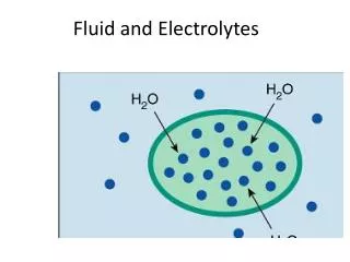
Electrolytes • ECF electrolytes • Sodium • Chloride • Bicarbonate • ICF electrolytes • Potassium • Phosphorus
Sodium • Dog 145 (140-155) mEq/L • Cat 156 (149-162) mEq/L • Horse 139 (132-146) mEq/L • Cattle 142 (132-152) mEq/L
Serum sodium concentration • Serum sodium concentration is an indication of the amount of sodium RELATIVE to the amount of water in ECF and provides no direct information about total body sodium content
Serum sodium concentration • Hypernatremia means hyperosmolality • Hyponatremia usually means hypoosmolality
Hypernatremia • Pure water loss • Hypotonic fluid loss • Gastrointestinal • Third space • Renal • Gain of impermeant solute
Hyponatremia • With hypervolemia • Severe liver disease, congestive heart failure, nephrotic syndrome • With normovolemia • Psychogenic polydipsia, anti-diuretic drugs, hypotonic fluids • With hypovolemia • GI loss, third space loss, hypoadrenocorticism, diuretics
Chloride • Dog 110 (105-115) mEq/L • Cat 120 (115-125) mEq/L • Horse 104 (99-109) mEq/L • Cattle 104 (97-111) mEq/L
Serum chloride concentration • Cl- and HCO3- are the main resorbable anions in renal tubular fluid and abnormalities in one often result in abnormalities of the other • Normal ratio of Na+ to Cl- in ECF is 1.3 to 1 and gain or loss of equal amounts of Na+ and Cl- will disturb this relationship. Only a gain or loss of 4 Na+ for every 3 Cl- would preserve this relationship
Hyperchloremia • Excessive loss of Na+ relative to Cl- (e.g. diarrhea) • Excessive gain of Cl- relative to Na+ (e.g. NH4Cl, 0.9% NaCl, hypertonic NaCl, salt poisoning) • Excessive Cl- retention by kidneys (e.g. compensation for chronic respiratory alkalosis)
Hypochloremia • Vomiting of stomach contents or sequestration of fluid in stomach • Diuretics (e.g. furosemide) • Compensation for chronic respiratory acidosis
Potassium • Dog 4.5 (3.5-5.5) mEq/L • Cat 4.5 (3.5-5.5) mEq/L • Horse 3.8 (2.6-5.0) mEq/L • Cattle 4.8 (3.9-5.8) mEq/L
Intake ECF Translocation ICF Excretion • Urinary (> 90%) • Fecal (< 10%) Potassium Balance
Translocation of potassium ECF ICF
Hyperkalemia • Increased intake (alone usually not sufficient to cause hyperkalemia if renal function adequate – unless iatrogenic) • Translocation (ICF to ECF) • Acute mineral acidosis, insulin deficiency • Decreased renal excretion • Urethral obstruction, uroabdomen, oligoanuric renal failure, hypoadrenocorticism, some drugs
Hypokalemia • Decreased intake (alone not usually sufficient to cause hypokalemia) • Translocation (ECF to ICF) • Alkalemia, insulin and glucose • Increased loss • GI, renal
Total CO2 or bicarbonate • Dog 21 (17-24) mEq/L • Cat 20 (17-24) mEq/L • Horse 27 (24-30) mEq/L • Cattle 25 (20-30) mEq/L
Total CO2 • Anaerobically measured, this includes HCO3-, dissolved CO2 and negligible amounts of carbamino-CO2, H2CO3, and CO3-2 • Aerobically measured, it is essentially equivalent to HCO3-
Total CO2 • Determination of total CO2 alone does not allow complete characterization of acid base disturbances • High total CO2 usually means metabolic alkalosis but compensation for respiratory acidosis could contribute • Low total CO2 usually means metabolic acidosis but compensation for respiratory alkalosis could contribute
Serum total calcium concentration • Dog 10.1 (9.0-11.3) mg/dL • Cat 9.2 (8.0-10.5) mg/dL • Horse 12.4 (11.2-13.6) mg/dL • Cattle 11.0 (9.7-12.4) mg/dL
Components of serum total calcium concentration Ionized Calcium (50%) Complexed Calcium (10%) Protein-bound Calcium (40%)
Normal serum calcium concentrations in dogs • Total: 9 to 11 mg/dl • Ionized: 5.1 to 5.7 mg/dl Routine serum biochemical profile returns serum total calcium concentration
Effect of acid base balance on serum calcium concentration • Acidosis tends to increase the ionized fraction and decrease the protein-bound fraction • Alkalosis tends to decrease the ionized fraction and increase the protein-bound fraction These results are due to the effects of acid base balance on the net charge of plasma proteins
5 5 1 1 4 Effect of hypoalbuminemia on serum calcium concentration Ionized Calcium (mg/dL) Complexed Calcium (mg/dL) 2 Protein-bound Calcium (mg/dL) Normal Hypoalbuminemia
Correction of hypocalcemia for hypoalbuminemia • Corrected Calcium = Calcium – Albumin + 3.5 • Works reasonably well in dogs • Unreliable in cats
5 2 4 Hypercalcemia in chronic renal failure (example) Ionized Calcium (mg/dL) 5 Complexed Calcium (mg/dL) 1 4 Protein-bound Calcium (mg/dL) CRF Normal
Hypercalcemia • Dehydration • Various malignancies • Hypoadrenocorticism • Renal failure • Hypervitaminosis D • Primary hyperparathyroidism
Hypercalcemia in horses with renal failure • May be related to fact that horses normally absorb large amounts of calcium from their GI tract and rely on their kidneys to excrete it (calcium carbonate crystals are common in horse urine)
Hypocalcemia • Hypoalbuminemia • Renal failure • Ethylene glycol poisoning • Eclampsia • Acute pancreatitis • Primary hypoparathyroidism
Phosphorus • Dog 4.2 (2.5-6.0) mg/dL • Cat 6.3 (4.5-8.1) mg/dL • Horse 4.3 (3.1-5.6) mg/dL • Cattle 6.0 (5.6-6.5) mg/dL
Serum phosphorus • Largely a mixture of H2PO4- and HPO4-2 • The net valence and number of mEq of phosphorus in ECF are influenced by pH hence it is easier to talk about phosphorus in terms of mMol or mg of elemental phosphorus
Hyperphosphatemia • Translocation (ICF to ECF) • Decreased renal excretion • Increased intake • Young growing animal
Hypophosphatemia • Translocation (ECF to ICF) • Decreased renal reabsorption • Decreased intestinal absorption Hypophosphatemia may occur in some horses with renal failure
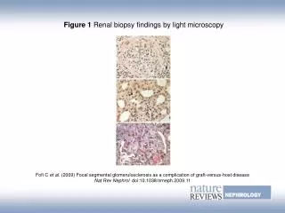
Renal Biopsy: Indications • Differentiation of renal diseases that may differ in their prognosis • Differentiation of ARF from CRF • Determination of status of basement membranes in ARF • Determination of response to therapy • Determination of progression of disease
Renal Biopsy: Contraindications • Coagulopathy • Severe hydronephrosis or perinephric pseudocyst • Renal or perirenal abscess • Pyonephrosis • Solitary kidney • Pyelonephritis • Renal neoplasia • Extremely small kidneys
Renal biopsy: General considerations • Adequate patient evaluation • Choice of technique • Choice of biopsy instrument • Direction of instrument into kidney • Hemostasis • Anesthesia
Pre-biopsy evaluation • Coagulation ability (role of buccal mucosal bleeding time) • IV catheter and fluid administration • PCV & TPP after fluids but before biopsy • Fluid diuresis
Methods of renal biopsy • Open surgical • True percutaneous • Keyhole • Laparoscopy • Ultrasound-guided (currently in use at OSU VTH) • Needle aspirate – NOT a biopsy!
What do those letters stand for? Renal biopsy: Keyhole technique
Post-biopsy evaluation • Fluid diuresis for 12 hours • Monitor PCV & TPP at appropriate intervals over 12 to 24 hours
Renal biopsy: Complications Microscopic hematuria vs macroscopic hematuria
Renal biopsy: Complications • Hemorrhage • Infarction • Hydronephrosis • Other extremely rare (e.g. infection, retention cyst, AV fistula, urine fistula)
Handling the biopsy • Avoid touching the biopsy specimen at all • Preservation of specimen • 10% buffered formalin for routine light microscopy and peroxidase-immunoperoxidase immunopathology • Michel’s medium for direct immunofluoresence • 2% glutaraldehyde for transmission electron microscopy