Download
1 / 14
140 likes | 170 Views
Explore the early stages of chick embryo development including node regression, notochord formation, and neurulation in the ectoderm. Learn about the organization of the body axis, development of extra-embryonic tissues, and the regression of Hensen’s Node. Dive into the typical vertebrate organization of the chick embryo and the formation of extra-embryonic membranes. Discover the process of mouse embryo development from fertilization to blastocyst formation and the implantation of ES cells for transgenesis.

E N D
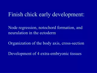
Finish chick early development:Node regression, notochord formation, and neurulation in the ectodermOrganization of the body axis, cross-sectionDevelopment of 4 extra-embryonic tissues
Regression of Hensen’s Node: 1) progressively laying down notochord toward the posterior tip and 2) progressively shutting down endomesoderm induction and waterfall ingression thru the streak.
N N N N Stage 6 Stage 7 Stage 4 18hr Stage 5 N N N Stage 8 Stage 7 N Stage 9 Stage 10 36hr Regression of Hensen’s Node
Typical vertebrate organization of the chick embryo(and >200 selector gene domains) “Somatic mesoderm” “Visceral mesoderm” Notochord **Or wait until the embryo lifts off the yolk sac
Amnion and chorion: contain ectoderm and somatic mesodermYolk sac and allantois: contain enododerm and visceral mesoderm
Start mouse development: fertilization to the 128 cell blastocyst
Human or mouse embryo: pre-implantation stages gene expressionstarts 2 2. Blastocyst cavity formation 3. Cell sorting (hypo vs. epi) 1. compaction 4. hatching Fallopian tube 128 cell Day 7-human Day 5.5-mouse
Mouse pre-implantation stages: compaction, apical-basal polarization, cleavage, blastocyst cavity formation 128-cell blastocyst compaction 128-cell blastocyst Compaction at the late 8-cell stage, With apical-basal polarization of cells
Oct4 gene expression in epiblast Human 128cell blastocyst before/during hatching “zona” Holding pipet Injection pipet Injection of ES cells into the blastocyst cavity Mouse embryonic stem cells (ES cells) from the inner cell mass, growing in a culture dish
Mouse transgenesis (insertion of a foreign gene into the mouse genome) Dr. Mario Capecchi-1984 Univ. of Utah (random) (e.g., GFP with promoter) FOSTERMOTHER non-homologous integration (2x) Green ES cells Gene for red fluorescent protein (RFP) Gene for green fluorescent protein (GFP) from jellyfish
Implantation of the human blastocyst in the uterine wall. Mouse implantation is also invasive, like human Syncytio- trophoblast Maternal tissue Uterine fluid