Download
1 / 57
590 likes | 810 Views
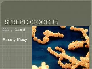
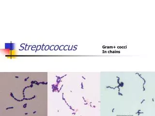
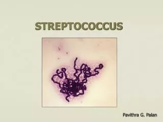
Streptococcus Species. Dr. Zaheer Ahmed Chaudhary Associate Professor Microbiology Department of Pathology. Introduction. One of the two main human pathogens. Gram positive cocci . Oval shaped. Arranged in chains and pairs. Catalase negative. Non motile. Facultative anaerobe.

E N D
Streptococcus Species Dr. Zaheer Ahmed Chaudhary Associate Professor Microbiology Department of Pathology
Introduction • One of the two main human pathogens. • Gram positive cocci. • Oval shaped. • Arranged in chains and pairs. • Catalase negative. • Non motile. • Facultative anaerobe. • Capsulated/ non capsulated.
Classification • Based on : • Serology: using Lancefields antibodies to cell wall carbohydrate. • Hemolysin test. • Biochemical tests.
Serology • It is based on C carbohydrate present in the cell wall and its specificity is determined by an amino sugar. • Antibodies to carbohydrate are detected and classified into A to U. • There are more than 20 groups in Lancefield grouping. • All Lancefield groups are pyogenic in nature.
Hemolysin test • There are three types of hemolysis on blood agar plate namely • Alpha hemolysis: Green zone around the bacterial colony which is due to incomplete hemolysis of RBCs in the agar. • Beta hemolysis: Clear zone of hemolysis around the bacterial colony of blood agar which is due to complete lysis of RBCs. It is produced by an enzyme hemolysinO & S.
Beta streptococci have two antigens in their cell wall. • C-carbohydrate • M protein M protein : is an important virulence factor and determines the type of group A, B streptococcus. It is antiphagocytic in nature. Approximately 80 serotypes are present on the basis of M protein. Some strains produce rheumatic fever and some cause kidney infections.
Gama Hemolysis: No hemolysis is produced on blood agar by the bacterial colonies.
Biochemical Tests • Following tests are done to classify different bacteria on the basis of chemical reaction. • CAMP Test – Streptococcus agalactae. • Growth inhibition to the bacitracin disc– Streptococcus pyogenes. • Sensitivity to optochin disc – Streptococcus Pneumoniae. • Bile solubility test – Streptococcus faecalis.
Medically important Streptococci • Streptococcus Pyogenes – Group A • Streptococcus agalactae – Group B • Streptococcus faecalis – Group D • Streptococcus bovis – Group D • Streptococcus Pneumoniae • Viridans group Streptococci
Diseases caused by Streptococcus • Strept. Pyogenes (Group A): produces bacterial diseases like cellulitis, pharyngitis, impetigo, necrotizing skin lesions and streptococcal toxic shock syndrome. • Two immunogenic diseases i.e Rheumatic fever and glomerulonephritis are also caused.
Streptococcus agalactae (Group B) : is the leading cause of neonatal sepsis and menigitis. • Streptococcus faecalis (Group D): causes hospital acquired urinary tract infection (UTI) and endocarditis.
Streptococcus Pneumoniae: causes pneumonia in young children and low immunity adults. • Viridans group Streptococci & Streptococcus bovis : common cause of endocarditis.
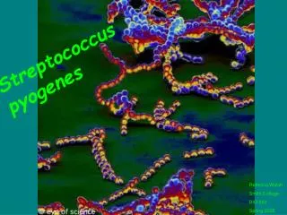
Beta Hemolytic Streptococci • Lancefield group A – U on the basis of antigenic differences in C- carbohydrates. • Group A : • Streptococcus pyogenes is the most important human pathogen causing sore throat and skin infections. • The organism adheres to the epithelium via pili covered by lipoteichoic acid and M proteins. • Growth is inhibited by bacitracin disc.
Group B: • Streptococcus agalactae colonizes in the genital tract of women, causes neonatal meningitis and sepsis. • Resistant to bacitricin.
Group D : Comprises of • Enterococci – Ent. faecalis, Ent. faecium • Non enterococci - Strept. bovis • They are normal flora of gut and can produce UTI, biliary and CVS infections. • Hardy organisms and can grow in saline and bile. • Non enterococci cause similar infections but the organisms are less hardy.
Group C , E, F, G, H, K-U : • Cause human diseases infrequently.
Transmission • Most Streptococci are part of the normal flora of skin, throat and intestine but produce diseases only when they gain entry to the blood circulation. • Viridance group and Strept. pneumonae are chiefly found in oropharaynx. • Strept. pyogenes is found on skin. • Streptagalactae is found in vagina and colon.
Pathogenesis • Pyogenic inflammation. • Exotoxin production. • Immunogenic.
Pyogenic Inflammation • M protein is the most important antiphagocytic factor. Group A produces 3 enzymes. • Hyaluronidase : It degrades hyaluronic acid which forms ground substance of subcutaneous tissue. It is also known as spreading factor since it facilitates the spread of infection to the tissue. • Streptokinase : It activates plasminogen to form plasmin which dissolves fibrin into clot, thrombi and emboli. It is used to lyse fibrin clots in coronary vessels in heart attack patients.
DNAse : Degrades DNA in exudates or nectrotic tissues. • Toxins: Various toxins are produced by streptococci. • Erythogenic toxins : It causes rash of scarlet fever. It acts as a super antigen.
Streptolysin O : It is hemolysin which is inactivated by oxidation and causes rheumatic fever. • Streptolysin S : It is oxygen stable and is not activated by oxidation. • Pyogenic exotoxin A : Causes toxic shock syndrome.
Pyogenic exotoxin B : Rapidly destroys the tissue and causes necrotizing fasciatis.
Clinical Finding • Group A causes pharangitis , sinusitis, mastoiditis, meningitis, TSS. It can also cause endometritis. • Group B causes neonatal sepsis, meningitis, pneumonia endocarditis and osteomylitis. • Group D causes UTI in hospitalized patients, indwelling catheters, pelvic infection.
Complications • Immunogenic response to streptococci can result in two forms : • Acute glomerulonephritis. • Rheumatic fever.
Acute glomerulonephritis: It occurs 2 to 3 weeks after the acute skin infection. Antigen-antibody complex gets deposited on glomerular basement membrane of the kidney leading to acute infection. This process can be stopped by avoiding colonization of skin by streptococcus group A.
Acute Rheumatic fever: It occurs two weeks after pharangitis. It is characterized by fever, migratory polyarthritis and carditis. Mitral and aortic valves are involved. Prevalent in children between the age of 5-15 years.
ASOT (Anti streptolysin O titer): • Rheumatic fever is due to immunological reaction between cross reacting antibodies to M protein of streptococcus A with antigens of joints , hearth and brain. • It can be prevented if treated within 8 days.
Lab Investigations • Clinical presentation. • Throat swab culture. • CAMP test. • Asculin test. • Serological tests (ASOT).
Treatment • Penicillin G or amoxicillin. • Erythromycin or Clindamycin in allergic patients.
Prevention • Prompt treatment to group A streptococcal infections. • Preventive benzyl penicillin every month. • Use of amoxycillin preoperatively. • All pregnant women should be screened for Group B streptococcus by vaginal and rectal cultures at 37 weeks gestation. • Penicillin is administered at the time of delivery. • No vaccine.
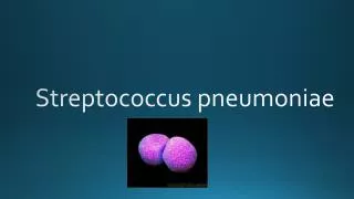
Introduction • Two types of streptococci which produce alpha hemolysis. • Streptococcus pneumoniae • Viridans Group Streptococcus
Streptococcus pneumoniae • Most common cause of community acquired pneumonia. • It produces following diseases namely bacteremia, meningitis, upper respiratory tract infection, otitis media, sinusitus, mastoiditis and conjuctivitis.
General Characteristics • Gram positive, lancet shaped cocci in pairs or short chains. • Tend to be oval with pointed ends. • Thick capsule around the organism. • Produce alpha hemolysis. • Lysed by bile salts. • Growth is inhibited by optochin disc on blood agar.
Capsule produces quellung reaction. • Capsule acts as virulance factor. • It interferes with phagocytosis. • C-substance in the cell wall reacts with normal serum proteins made by liver called CRP. • It is increased in acute infection, 1000 folds and is used to monitor the progress of the disease.
Transmission • 5-50% of normal healthy people harbor virulent organism in oropharynx. • It is not a communicable disease. • Disease incidence increases with predisposing factors which include : • Alcohol or drug intoxication. • Abnormality in the respiratory system. • Pulmonary congestion. • Splenectomy
Pathogenesis • Pneumococci produces IgA protease which enhances the ability of the organism to colonize in upper respiratory tract. • It multiples in the tissue and causes inflammation. • Polysacchride capsule is the main virulence factor.
Lipoteichoic acid activates the complement and induces inflammatory cytokines production.
Clinical findings • Sudden chills, fever, cough and pleuretic pain. • Sputum is red / brown color. • It is prominent cause of ear infection sinus, eyes, bronchioles and sepsis in immunocompromised patients.
Lab Diagnosis • Gram smear - Lancet shaped Gram positive diplococci. • Smear Culture - Positive in 15-25% cases. Draughtsmen colonies on blood agar. • Quellung Reaction. • Bile soluble • Alpha hemolytic colonies on blood agar. • Growth inhibited by optochin. • CSF culture.