Download
1 / 12
120 likes | 132 Views
Lupus erythematosus. Definition. Lupus erythematosus (LE) is an autoimmune disease of unknown cause involving the skin and/or other organs. LE is the designation of a spectrum of diseases that are linked by distinct clinical findings and distinct pattern of polyclonal B cell immunity.

E N D
Definition • Lupus erythematosus (LE) is an autoimmune disease of unknown cause involving the skin and/or other organs. • LE is the designation of a spectrum of diseases that are linked by distinct clinical findings and distinct pattern of polyclonal B cell immunity. • It ranges from life-threatening manifestations of systemic LE (SLE) to the limited and exclusive skin involvement in chronic cutaneous LE (CCLE). • Lupus erythematosus is a complex and highly variable disease in which a combination of genetic, immunologic and environmental factors acts in concert with the production of the disorder.
LE immunologic factors • A wide range of immune abnormalities may contribute to autoantibody production: • polyclonal B-cell activation, • molecular mimicry and antibody cross reactivity, • loss of T-cell tolerance, • abnormal T-cell help, • cytokine abnormalities (increased production of IL-1, IL-4, IL-6 and IFN gamma), etc.
LE immunologic factors • Auto antibodies can induce tissue damage in LE by two mechanisms: • they can bind directly to cells, resulting in type II immunologycally mediated tissue damage; • they can bind to circulating antigens with formation of immune complexes; leading to type III immunologically mediated tissue damage.
LE auto-antibodies • Antinuclear antibodies (ANA); • Antibodies to double-stranded DNA (anti ds DNA); • Antibodies to single-stranded DNA (anti ss DNA); • Anti-SM antibody; • Anti-Ro antibodies (anti SS-A); • Anti-La antibodies (anti SS-B); • Anti-ribonucleoproteins (anti RNP)
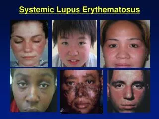
LE Classification • Systemic LE (SLE) • acute cutaneous LE (ACLE): malar rash, discoid rash, photosensitivity, oral ulcers • arthritis: nonerosive involving two or more peripheral joints • serositis: pleuritis; pericarditis • renal disorder: persistent proteinuria; cellular casts • neurologic disorder: seizures; psychosis • hematologic disorder: hemolytic anemia; leukopenia; lymphopenia; thrombocytopenia • immunologic disorder: anti-DNA, anti-SM and antiphospholipid antibodies • antinuclear antibody: abnormal titer of antinuclear antibodies • Cutaneous LE (CLE) • subacute cutaneous LE (SCLE) • chronic cutaneous LE (CCLE)
Classification of LE skin lesions • I. LE-specific skin disease (CLE) A. Acute cutaneous LE (ACLE) 1.Localized ACLE (malar rash, butterfly rash) 2.Generalized ACLE (maculopapular lupus rash, malar rash, photosensitive lupus dermatitis) B. Subacute cutaneous LE (SCLE) 1. Annular SCLE 2. Papulosquamous SCLE C. Chronic cutaneous LE (CCLE) 1. Classic discoid LE (DLE) 2. Disseminated DLE 3. Superficial LE (Besnier’s centrifugal erythema) 4. LE profundus (chronic lupus panniculitis) 5. Mucosal DLE (oral DLE, conjunctival DLE) 6. Lupus tumidus (urticarial plaque of LE) 7. Chilblains LE 8. Lichenoid DLE (LE / lichen planus overlap). • II. LE-nespecific skin disease These range from necrotizing and urticarial vasculitis to livedo reticularis, Raynaud’s phenomenon, dermal muconosis, and bullous lesions in LE.
Discoid Chronic Cutaneous LE • The basic symptoms: erythematic plaques, hyperkeratotic plaques and atrophy. • Early lesions are usually confined to head and neck and appearing as inflammatory, erythematous, edematous and scaling macules or papules, with few mm in d, which spread centrifugally into larger plaques. The scales are adherent, with horny plugs in dilated pilosebaceous canals. • The removal of the scale (Besnier-Mescerski sign) demonstrates a characteristic „carpet tack” appearance corresponding to patulous and plugged follicular orifices. • The face is most commonly affected. Other sites are: scalp, neck, ears, hands, rarely arms, legs and trunk. Permanent scarring alopecia occurs in the scalp lesions. • Nail changes are subungual hyperkeratosis, longitudinal striae, and red-blue colorings of the nail plate. • Mucous membrane lesions are erythematous patches, hyperkeratotic plaques, leucoplakia and ulcerations, commonly located on the inner cheeks, tongue, lips and palate.
Discoid Chronic Cutaneous LE • The secondary symptoms: • pigmentary disturbances: hyperpigmentation and hypopigmentation • telangiectases • infiltration are frequently associated. • The evolution with scarring is common.
Discoid Chronic Cutaneous LE Atypical forms • Warty type, with marked hyperkeratosis and the formation of a warty plaque or nodule. Common sites are nose, temples, ears, scalp, dorsal hands, but also palms and soles; • Tumidus form, with swollen, warm and tense plaques on the check, on a limb; • Chilblain lupus, with perniotic lesions on the toes and fingers, but also on the heels, calves, knees, elbows, nose and ears. • Lupus panniculitis (profundus): movable subcutaneous nodule on the arms, forehead, cheecks, chin, back, buttocks, thighs, scalp, breasts, eyelids etc. • LE centrifugal erythema of Besnier – superficial form.
LE Laboratory Investigations • LE cell test is positive in over 80% of patients with SLE; • Antinuclear antibodies test (ANA test) – 95-100% in SLE; • Antibodies to double-stranded DNA (anti ds DNA); • Anti-SM antibody; • Anti-Ro antibodies (anti SS-A); • Anti-La antibodies (anti SS-B); • Serum complement level is low; • Lupus band test: direct immunofluorecence evidencing IgG and IgM and complement (C3) in a continuous granular line or band along the dermo-epidermal junction – in 90% of active lesions. • Histology: vacuolar degeneration of epidermal basal cells, hyperkeratozis, atrophy of the epidermis, follicular plugging, papillary dermal edema, a perivascular mononuclear infiltrate and extravasation of erythrocytes.
TreatmentCutaneous LE • Topical therapy • topical corticosteroids: potent or superpotent are effective (methylprednisolone aceponate, mometasone furoate, hydrocortisone butirate, betamethasone dipropionate, flucasone propionate, flucinonide, halcinonide, amcinonide, clobetasol dipropionate, halbetasol propionate etc); • sun-screens (SPF 30); • Systemic treatment • antimalarials: chloroquine sulphate, hydroxychloroquine; • corticosteroids; • thalidomide; • retinoids; • dapsone; • clofazimine