Download
1 / 69
750 likes | 2.35k Views
Inflammation and Repair. Learning Objectives:. Define inflammation, its causes and clinical appearance. Describe the sequence of vascular changes in acute inflammation ( vasodilation , increased permeability) and their purpose. Know the mechanisms of increased vascular permeability.

E N D
Learning Objectives: • Define inflammation, its causes and clinical appearance. • Describe the sequence of vascular changes in acute inflammation (vasodilation, increased permeability) and their purpose. • Know the mechanisms of increased vascular permeability. • Define the terms edema, transudate, and exudate.
Learning Objectives: • Define inflammation, its causes and clinical appearance. • Describe the sequence of vascular changes in acute inflammation (vasodilation, increased permeability) and their purpose. • Know the mechanisms of increased vascular permeability. • Define the terms edema, transudate, and exudate.
Inflammation • Inflammation is a protective response intended to eliminate the initial cause of cell injury as well as the necrotic cells and tissues resulting from the original insult • The reaction of vascularized living tissue to local injury.
How to accomplishes protective mission? • Inflammation serves to destroy, dilute or isolate the injurious agent (microbes, toxins) and eliminate the necrotic cells and tissues. • Inflammation is part of a broader protective response (innate immunity ) • It starts a series of events which leads as far as possible to the healing and reconstitution of the damaged tissue.
Inflammation • During repair, the injured tissue is replaced by : • Regeneration of native parenchyma cells • Filling of the defect by fibroblastic tissue or both • Inflammation and repair are protective response
Can Inflammation cause considerable harm to the body? They may induce harm e.g. anaphylactic reaction rheumatoid arthritis atherosclerosis pericarditis
HOW? • The components of the inflammatory reaction that destroy and eliminate microbes and dead tissues are capable of also injuring normal tissues. • This may accompany • entirely normal, beneficial inflammatory reactions, (e.g., when the infection is severe), • prolonged (e.g., when the eliciting agent resists eradication) • inappropriate (e.g., when it is directed against self-antigens in autoimmune diseases) • against usually harmless environmental antigens (allergic disorders)
Then what happen? • Inflammation is terminated when the offending agent is eliminated and the secreted mediators are broken down or dissipated. • There are active anti-inflammatory mechanisms that serve to control the response and prevent it from causing excessive damage to the host.
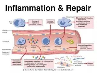
What are the cells and molecules that play important roles in inflammation? • blood leukocytes • plasma proteins • cells of vascular walls • cells of the surrounding connective tissue • extracellular matrix (ECM) of the surrounding connective tissue
The connective tissue cells • The circulating cells: The extracellular matrix
Cells and molecules that play important roles in inflammation
Tissues and cells involved in inflammatory response : • The fluid and proteins of plasma, circulating cells, blood vessels and connective tissue • The circulating cells: neutrophils, monocytes, eosinophils, lymphocytes, basophils, and platelets. • The connective tissue cells are the mast cells, the connective tissue fibroblasts, resident macrophages and lymphocytes. • The extracellular matrix, consists of the structural fibrous proteins (collagen, elastin), adhesive glycoproteins (fibronectin, laminin, nonfibrillar collagen, tenascin, and others), and proteoglycans
Inflammation • Inflammation is divided into: • Acute inflammation. • Chronic inflammation.
Acute inflammation Chronic inflammation • rapid in onset (seconds or minutes) • relatively short duration, lasting for minutes, several hours, or a few days • its main characteristics: • the exudation of fluid and plasma proteins (edema) • the emigration of leukocytes, predominantly neutrophils. • is of longer duration • associated histologically with the presence of lymphocytes and macrophages, the proliferation of blood vessels, fibrosis, and tissue necrosis. • Less uniform.
Source of chemical mediators in inflammation • Phagocytes and other host cells • Leukocyte • Endothelium • Mast cell • Plasma proteins
What is the action of chemical mediators in inflammation? • Some of mediators act on small blood vessels • Promote the efflux of plasma • Recruitment of circulating leukocytes to the site where the offending agent is located
Acute Inflammation • Local clinical signs of acute inflammation : • Heat • Redness • Swelling • Pain • Loss of function
What are the steps of the inflammatory response which can be remembered as the five Rs?
Steps of the inflammatory response (1) Recognition of the injurious agent (2) Recruitment of leukocytes (3) Removal of the agent (4) Regulation (control) of the response (5) Resolution
Learning Objectives: • Define inflammation, its causes and clinical appearance. • Describe the sequence of vascular changes in acute inflammation (vasodilation, increased permeability) and their purpose. • Know the mechanisms of increased vascular permeability. • Define the terms edema, transudate, and exudate.
Components of acute inflammation Vascular changes Cellular events • Vasodilation: alterations in vessel caliber resulting in increased blood flow • Increased vascular permeability: permit plasma proteins to leave the circulation • Emigration of the leukocytes from the microcirculation and accumulation in the focus of injury • Principal leukocytes in acute inflammation are neutrophils (polymorphonuclear leukocytes).
Causes of Acute Inflammation • Triggered by a variety of stimuli: • Infections (bacterial, viral, parasitic) and microbial toxins • Trauma (blunt and penetrating) • Physical and chemical agents (thermal injury, e.g., burns or frostbite; irradiation; some environmental chemicals) • Tissue necrosis (from any cause) • Foreign bodies (splinters, dirt, sutures) • Immune reactions (also called hypersensitivity reactions)
In summary • Acute inflammation is a rapid response to an injurious agent that serves to deliver mediators of host defense-leukocytes and plasma proteins-to the site of injury. • Acute inflammation has three main events: (1) Hemodynamic changes (alterations in vascular caliber that lead to an increase in blood flow) (2) Increased vascular permeability (structural changes in the microvasculature that permit plasma proteins and leukocytes to leave the circulation) (3) Emigration of the leukocytes from the microcirculation (their accumulation in the focus of injury, and their activation to eliminate the offending agent)
Vascular Changes (1) Hemodynamic changes (alterations in vascular caliber that lead to an increase in blood flow) (2) Increased vascular permeability
What are the different phases of changes in Vascular Caliber and Flow?
Phases of changes in Vascular Caliber and Flow • 1. Transient vasoconstriction of arterioles - It disappears within 3-5 seconds in mild injuries - It may last several minutes in more severe injury (burn)
Phases of changes in Vascular Caliber and Flow • 2.Vasodilatation: It involves the arterioles and then results in opening of new microvasculature beds in the area thus leading to increasing blood flow (early hemodynamic change and the cause of inflammation heat and redness). • Increased blood volume lead to increased local hydrostatic pressure leading to transudation of protein -poor fluid into the extravascular space.
Phases of changes in Vascular Caliber and Flow • 3.Slowing of the circulation due to increased permeability of the microvasculature, this leads to outpouring of protein-rich fluid in the extravascular tissues. This results in concentration of the red cells in small vessels and increased viscosity of the blood leadind to stasis (stasis: slow circulation due to dilated small vessels packed with red cells).
Phases of changes in Vascular Caliber and Flow • 4. persistence of stasis leads to peripheral orientation of leukocytes (mainly neutrophils) along the vascular endothelium [leukocytic margination], then they migrate through the vascular wall into the interstitial tissue [emigration].
A 72-year-old man with severe emphysema has had worsening right ventricular failure for the past 5 years. He has had fever and increasing dyspnea for the past 4 days. A chest radiograph shows an accumulation of fluid in the pleural spaces. Fluid obtained by thoracentesis has a specific gravity of 1.030 and contains degenerating neutrophils. The most likely cause of this fluid accumulation is an increase in which of the following mechanisms? (A) Colloid osmotic pressure (B) Lymphatic pressure (C) Vascular permeability (D) Renal retention of sodium and water (E) Leukocyticdiapedesis
Increased Vascular Permeability • A hallmark of acute inflammation(escape of a protein-rich fluid). • It affects small & medium size venules, through gaps between endothelial cells
Marked outflow of fluid and its accumulation in the interstitial tissue: • The loss of protein from the plasma reduces the intravascular osmotic pressure and increases the osmotic pressure of the interstitial fluid • the increased hydrostatic pressure • The net increase of extravascular fluid results in edema Slide 3.3
Learning Objectives: • Define inflammation, its causes and clinical appearance. • Describe the sequence of vascular changes in acute inflammation (vasodilation, increased permeability) and their purpose. • Know the mechanisms of increased vascular permeability. • Define the terms edema, transudate, and exudate.
What is the edema? • Edema • denotes an excess of fluid in the interstitial or serous cavities • it can be either an exudate or a transudate.
Exudate • An inflammatory extravascular fluid that has a high protein concentration, cellular debris, and a specific gravity above 1.020. • It implies significant alteration in the normal permeability of small blood vessels in the area of injury. Transudate • A fluid with low protein content (most of which is albumin) and a specific gravity of less than 1.012. • It is essentially an ultrafiltrate of blood plasma that results from osmotic or hydrostatic imbalance • No an increase in vascular permeability. Pus (a purulent exudate) • is an inflammatory exudate rich in leukocytes (mostly neutrophils), the debris of dead cells and microbes.