Download
1 / 2
20 likes | 125 Views
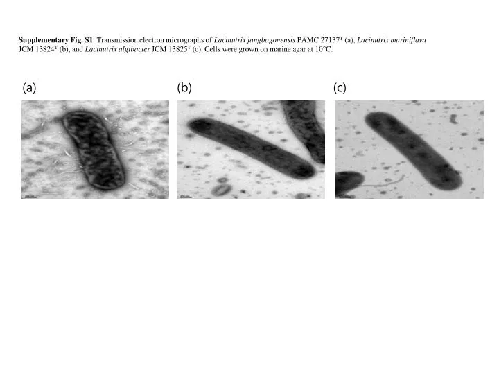
Supplementary Fig. S1. Transmission electron micrographs of Lacinutrix jangbogonensis PAMC 27137 T (a), Lacinutrix mariniflava JCM 13824 T (b), and Lacinutrix algibacter JCM 13825 T (c). Cells were grown on marine agar at 10 C. (a). (b). (c).

E N D
Supplementary Fig. S1. Transmission electron micrographs of Lacinutrixjangbogonensis PAMC 27137T (a), LacinutrixmariniflavaJCM 13824T (b), andLacinutrixalgibacterJCM 13825T (c). Cells were grown on marine agar at 10C. (a) (b) (c)
Supplementary Fig. S2. Two-dimensional thin layer chromatograms of the total polar lipids of Lacinutrixjangbogonensis PAMC 27137T (a), LacinutrixmariniflavaJCM 13824T (b), andLacinutrixalgibacterJCM 13825T (c), after spraying the plate with molybdatophosphoric acid. PE, phosphatidylethanolamine; AL1-AL2, unidentified aminolipids; APL, unidentified aminophospholipid; L1–L6, unidentified lipids. Lipids were identified as phospho- or amino- by spraying with molybdenum blue and ninhydrin, respectively. L1 L2 PE APL (a) (b) (c) AL1 L3 L4 L5 L1 L6 L2 PE AL2 AL1 L3 L4 L5 L1 L2 PE AL1 L3 L4