Download
1 / 44
440 likes | 722 Views
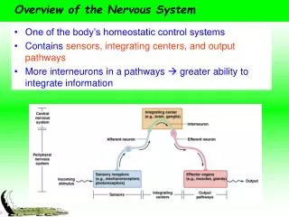
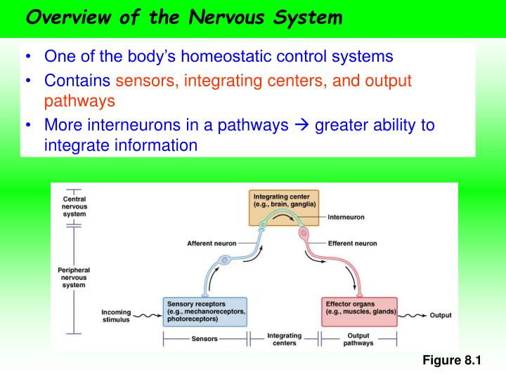
Overview of the Nervous System. One of the body’s homeostatic control systems Contains sensors, integrating centers, and output pathways More interneurons in a pathways greater ability to integrate information. Figure 8.1. Cnidarians.

E N D
Overview of the Nervous System • One of the body’s homeostatic control systems • Contains sensors, integrating centers, and output pathways • More interneurons in a pathways greater ability to integrate information Figure 8.1
Cnidarians • Most nervous systems are organized into three functional divisions • Cnidarians are an exception • Their nervous system is an interconnected web or nerve net • Neurons are not specialized into different divisions • Neurons are functionally bipolar and impulses radiate out from the stimulus • Can still perform complex behaviors
Nervous System Terms • Bilaterally symmetrical – anterior and posterior end and a right and left side • Cephalization - sense organs are concentrated at the anterior end • Brain – a complex integrating center made up of clusters of ganglia • Ganglia– groupings of neuronal cell bodies • Nuclei– groupings or neuronal cell bodies within the brain • Tracts – groupings of axons within the brain • Nerves – axons of afferent and efferent neurons
Structure of a Nerve • Parallel bundles of myelinated and unmyelinated axons enclosed in several layers of connective tissue • Endoneurium • Perineurium • Epineurium • Fasicles – bundle of axons • Mixed nerves – contain both afferent and efferent neurons Figure 8.3
Nervous Systems Across Animal Groups • Cephalization occurs in most animals and becomes more apparent in more complex nervous systems • Cnidarians and Echinoderms are exceptions • Organisms with more complex nervous systems have more neurons
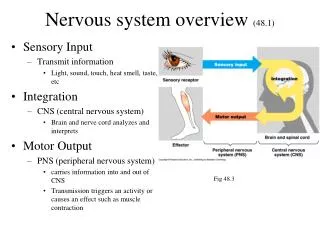
The Vertebrate Central Nervous System • Among the most highly cephalized animals • Unique in having a hollow dorsal nerve cord • Portion of nervous system is encased within cartilage or bone • Central nervous system (CNS) – brain and spinal cord • Peripheral nervous system (PNS) – rest of the nervous system Figure 8.5a
Cranial and Spinal Nerves • Cranial nerves • Exit directly from the braincase • 13 pairs (labeled with roman numerals) • Some are afferent and some are efferent • Spinal nerves • Emerge from the spinal cord • Named based on the region of the spine where they originate
Gray and White Matter • Brain and spinal cord contain two types of tissue • Gray matter – neuronal cell bodies • White matter – bundles of axons and their myelin sheaths • Spinal chord white matter is on the surface and gray matter is inside (opposite for cerebral cortex) Figure 8.5b
The CNS is Isolated • Meninges– layers of connective tissue that surround the brain and spinal cord • Number of layers vary across taxa (fish have one, mammals have three) • Cerebral spinal fluid (CSF) fills the space within the meninges and acts as a shock absorber • Blood-brain barrier – tight junctions in brain capillaries prevent material from leaking out of the bloodstream and into the CNS Figure 8.6
The Vertebrate Brain • The brain is an extension of the spinal cord • It is hollow inside and central cavities called ventricles contains CSF • Three main regions • Rhombencephalon (hindbrain) • Reflexes and involuntary behaviors • Mesencephalon (midbrain) • Coordination of sensory information • Relay center in mammals • Prosencephalon (forebrain) • Integration of olfactory information with other senses • Regulation of body temperature, reproduction, eating, emotion • Learning and memory in mammals
Brain Size • Most groups have the same major brain structures, although these structures vary in relative size Figure 8.9
The Parts of the Mammalian Brain Table 8.2
Hindbrain • Three regions • Pons – located above the medulla • Pathway between the medulla, the cerebellum, and the forebrain • Controls alertness and initiates sleep and dreaming • Cerebellum – two hemispheres at the back of the brain • Responsible for motor coordination • Contains half of the neurons in the brain • Medulla oblongata – located at the top of the spinal cord • Regulates breathing, heart rate, diameter of blood vessels, and blood pressure • Contain pathways between the spinal cord and the brain • Many cross over (e.g., left to right)
Midbrain • Primary center for coordinating and initiating behavioral responses in fish and amphibians • Size and function reduced in mammals • Primarily serves as a relay center • Sometimes grouped with the pons and medulla and termed the brainstem
Forebrain • Involved in processing and integrating sensory information, and in coordinating behavior • Main regions • Cerebrum • Thalamus • Epithalamus • Hypothalamus
Cerebrum • Outer layer is the cortex • Divided into two cerebral hemispheres • Left side controls the right side of the body • Right side controls the left side of the body • Connected by the corpus callosum
Cortex • Integrates and interprets sensory information and initiates voluntary movements • Has taken over many of the midbrain functions in lower vertebrates • Six layers • Isocortex (outer layer) is necessary for cognition and higher brain functions • More folded in more advanced mammals • Gyri – folds • Sulci – grooves
Cortical Lobes • Based on the names of the overlying bones or function Figure 8.14
Cortical Topology • Each part of the cortex corresponds to the specific part of the body that it governs • The areas devoted to various parts of the body are disproportionate Figure 8.5
Hypothalamus • Located at the base of the forebrain • Maintains homeostasis • Interacts with the autonomic nervous system • Regulates secretion of pituitary hormones
Limbic System • A network of connected structures that lie between the cortex and the rest of the brain • Influences emotions, motivation, and memory • Sometimes called the “emotional brain” • Includes the hypothalamus and other parts • Amygdala – aggression and fear responses • Hippocampus– converts short-term memory to long-term memory • Olfactory bulbs – sense of smell Figure 8.11
Thalamus • Large grouping of gray matter above the hypothalamus • Part of the reticular formation • Receives input from the limbic system and all senses except olfaction • Relays information to the cortex • Acts as a filter
Epithalamus • Located above the thalamus • Contains • Habenular nuclei – communicates with the tegmentum of the midbrain • Pineal complex – Establishes circadian rhythms and secretes melatonin
Peripheral Nervous System Divisions Figure 8.16
Autonomic Pathways • Involved in homeostasis • “Involuntary nervous system” • Systems • Sympathetic • Most active during periods of stress or physical activity • “Fight-or-flight” system • Parasympathetic • Most active during periods of rest • “Resting and digesting” system • Enteric • Independent of other two systems • Affects digestion by innervating the GI tract, pancreas, and gall bladder
Maintaining Homeostasis • Balancing of the sympathetic and parasympathetic systems • Three features of maintaining homeostasis • Dual innervation – most internal organs receive input from both systems • Antagonistic action – one system stimulates while the other inhibits • Basal tone – Even under resting conditions autonomic neurons produce APs
Dual Innervation Figure 8.17
Antagonistic Action Table 8.3
Similarities in Autonomic Pathways • Pathways contain two neurons in series • Preganglionic– may synapse with many postganglionic neurons and intrinsic neurons • Postganglionic – release neurotransmitter at the effector from varicosities • These neurons synapse with each other in the autonomic ganglia Figure 8.18
Differences in Autonomic Pathways • Differences between the sympathetic (S) and parasympathetic (PS) branches • Preganglionic cell body location • S: thoracic and lumbar regions of the spinal cord • PS: hindbrain and sacral region of the spinal cord • Ganglia location • S: chain that runs close to the spinal cord • PS: close to the effector • Number of postganglionic neurons that synapse with a single preganglionic neuron • S: 10 or more • P: three or less
Differences in Autonomic Pathways, Cont. • Type of neurotransmitter released at the effector Figure 8.19
Only Sympathetic Innervation • Some effectors receive only sympathetic innervation • Adrenal medulla – modified postganglionic neuron • Sweat glands • Arrector pili muscles in the skin • Kidneys • Most blood vessels Figure 8.20
Sympathetic vs. Parasympathetic Systems Table 8.4
Regulation of the Autonomic System Figure 8.21
Reflex Arcs • Most autonomic changes occur via simple neural circuits that do not involve conscious centers of the brain Figure 8.22
Somatic Motor Pathways • Control skeletal muscle • Usually under conscious control • The “Voluntary nervous system” • Some pathways are not under conscious control, e.g., knee-jerk reflex
Somatic Pathway Characteristics • Control only one type of effector, skeletal muscle • Cell bodies are located in the CNS • Monosynaptic, therefore very long • Axons split into a cluster of axon terminals at the neuromuscular junction • Synaptic cleft between the motor neuron and the muscle is very narrow • Release the neurotransmitter acetylcholine • Effect on the muscle is always excitatory
Reflex Arcs • Least complex integrated responses • Can involve as few as two neurons (monosynaptic) or more than two (polysynaptic) Figures 8.23 & 8.25
Reflex Arcs, Cont. • Can be arranged in two ways • Convergence – allows spatial summation • Divergence – can amplify signals Figure 8.24
Learning and Memory • Most animals can form memories and learn due to the plasticity of the nervous system • Learning – process of acquiring new information • Memory – retention and retrieval of information • Plasticity – ability to change both synaptic connections and functional properties of neurons in response to stimuli
Invertebrate Learning and Memory • Well studied in the sea slug, Aplysia (20,000 neurons) • Habituation – decline in response to a stimulus due to repeated exposure • Allows animal to ignore unimportant stimuli and focus on novel stimuli • Occurs because of changes in the presynaptic axon terminal • Inactivation of Ca2+ channels neurotransmitter release Figure 8.29
Invertebrate Learning and Memory, Cont. • Sensitization – increase in the response to a gentle stimulus after exposure to a strong stimulus • Occurs because of changes in the presynaptic axon terminal • Ca2+ entry neurotransmitter release • Involves a secondary circuit • Releases serotonin binds to G-protein-coupled receptors cascade of reactions inactivation of K+ channels AP duration Ca2+ influx neurotransmitter release
Serotonin Effects Figure 8.31
Memory in Mammals • The hippocampus is involved in long-term memory, but the memories are stored elsewhere • Long-term potentiation – repetitive stimulation of hippocampal tissue leads to an increase in the response of the postsynaptic neuron