Download
1 / 27
390 likes | 1.16k Views
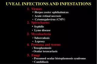
UVEAL INFECTIONS AND INFESTATIONS. 1. Viruses. Herpes zoster ophthalmicus Acute retinal necrosis Cytomegalovirus (CMV). 2. Spirochaetes. Syphilis Lyme disease. 3. Mycobacteria. Tuberculosis Leprosy. 4. Protozoa and worms. Toxoplasmosis Ocular toxocariasis. 5. Fungi.

E N D
UVEAL INFECTIONS AND INFESTATIONS 1. Viruses • Herpes zoster ophthalmicus • Acute retinal necrosis • Cytomegalovirus (CMV) 2. Spirochaetes • Syphilis • Lyme disease 3. Mycobacteria • Tuberculosis • Leprosy 4. Protozoa and worms • Toxoplasmosis • Ocular toxocariasis 5. Fungi • Presumed ocular histoplasmosis syndrome • Candidiasis
Herpes zoster ophthalmicus • Iritis in 40% of cases • Within 3 weeks of onset of rash Small-medium KP Iris atrophy - 20% Particularly if external nasal branch involved - Hutchinson sign
Acute retinal necrosis • Affects healthy individuals (bilateral in 30-50 %) • Herpes simplex in young patients • Herpes zoster in older patients • Vitritis and anterior uveitis • Peripheral vasculitis • Deep, multifocal, yellow, • necrotic infiltrates • Confluence but sparing of • posterior pole until late • Residual RPE atrophy • after 4-12 weeks
Acute retinal necrosis Treatment options • Systemic aciclovir, • steroids, aspirin • Laser photocoagulation • to limit progression Complications • Retinal detachment • Ischaemic optic neuropathy
Acquired immune deficiency syndrome (AIDS) Opportunistic infections Candidiasis • Toxoplasmosis • Atypical mycobacterium • Cytomegalovirus • Cryptococcus • Pneumocystis carinii • pneumonia Neoplasms • Kaposi sarcoma • Lymphoma
Anterior features Eyelid Kaposi sarcoma Conjunctival Kaposi sarcoma Multiple molluscum contagiosum Severe herpes zoster ophthalmicus Peripheral herpes simplex keratitis Microsporidial keratitis
HIV retinal microangiopathy • In 66% of AIDS • In 40% of AIDS-related complex • In 1% of asymptomatic HIV • infection • Transient cotton-wool spots • Occasionally haemorrhages
Indolent CMV retinitis • Slow progression • Frequently starts in periphery • No vasculitis • Granular opacification • Mild vitritis
Fulminating CMV retinitis • Dense, white, confluent opacification • May be associated with venous • sheathing • Frequently along vascular arcades • Mild vitritis • Associated haemorrhages
Progression of CMV retinitis Optic nerve head involvement ‘Brushfire-like’ extension along course of retinal blood vessels Extensive retinal atrophy Atrophy and retinal detachment
Treatment of CMV retinitis Ganciclovir Foscarnet i.v. • Systemic - initially i.v. then oral Cidofovir i.v. • Intravitreal - injections or • slow-release devices Signs of regression • Fewer haemorrhages • Less opacification • Diffuse atrophic and • pigmentary changes
Other fundus lesions in AIDS Progressive outer retinal necrosis Atypical toxoplasmosis Choroidal pneumocytosis Cryptococcal choroiditis Candidiasis Large cell lymphoma
Syphilis • Infection with spirochaete Treponema pallidum • Uveitis may occur during secondary and tertiary stages • Uncommon, bilateral in 50% Iridocyclitis Initially may be associated with dilated vessels (roseolae) Becomes chronic unless treated
Posterior syphilitic uveitis Multifocal chorioretinitis Unifocal chorioretinitis • May be bilateral • May be bilateral • Frequently juxtapapillary • or central • Residual choroidal atrophy • and RPE changes Acute neuroretinitis Inactive neuroretinitis • Usually unilateral • Optic atrophy, vascular • non-perfusion and RPE • changes • Disc oedema, macular • star and cotton wool spots
Lyme disease • Infection with Borrelia burgdorferi • Transmitted through bite of tick Ixodes sp. • Early and late manifestations Cardiac conduction defects Skin rash (erythema migrans) CNS lesions Mono-arthritis
Ocular features Acute conjunctivitis Punctate subepithelial keratitis Anterior uveitis Papilloedema Intermediate uveitis Neuroretinitis
Tuberculosis • Infection with human (M. tuberculosis) or bovine (M. bovis) • Uveitis is uncommon and occurs during post-primary stage • Positive skin test • Lung cavitation • Negative chest X-ray does not • exclude TB • Useful in diagnosis • of extrathoracic TB
Tuberculosis uveitis Chronic granulomatous iridocyclitis Mutton fat KP Koeppe nodules Busacca nodules Posterior uveitis Large solitary choroidal granuloma Choroiditis - unifocal or multifocal Retinal periphlebitis
Leprosy • Infection with M. leprae • Two types of leprosy - lepromatous and tuberculoid • Affinity for skin, peripheral nerves and eye Neurological involvement Severe corneal scarring Madarosis and skin involvement Lagophthalmos
Chronic lepromatous iritis Caused by invasion of anterior uvea by bacilli • Initially small, peripupillary, • glistening ‘iris pearls’ • Eventual iris atrophy, miosis • and cataract • Pearls enlarge and drop • into anterior chamber
Toxoplasmosis • Intracellular protozoan Toxoplasma gondii • Cat is definitive host • Other animals and humans are intermediate hosts Life cycles
Congenital systemic involvement Severity of involvement of fetus depends on duration of gestation at time of maternal infestation Infestation during late pregnancy may cause hydrocephalus Chorioretinal scarring at macula which may be bilateral
Toxoplasma retinitis • Recurrence of healed congenital lesion • Usually between ages 10-35 years. Vitritis may be severe Unifocal retinitis adjacent to old scar - heals within 1 to 4 months - ‘headlight in fog’
Treatment of toxoplasma retinitis Indications • Lesions at posterior pole, near optic disc or major blood vessel • Very severe vitritis • AIDS patients irrespective of location or severity Drugs 1. Systemic steroids 2. Clindamycin 3. Sulphonamides 4. Pyrimethamide 5. Co-trimoxazole 6. Azithromycin 7. Atovaquone
Ocular toxocariasis Always unilateral Chronic endophthalmitis Posterior pole granuloma Presents between 2 to 9 years. with leukocoria or strabismus Presents between 6 to 14 years. with visual loss Optic nerve granuloma Peripheral granuloma Presents during adolescence or adult life with visual loss Presents between 6 to14 years. with visual loss
Presumed ocular histoplasmosis syndrome • Fungal infection - Histoplasma capsulatum • Vitreous is never involved Peripapillary atrophy Atrophic ‘histo’ spots Choroidal neovascularization Peripheral streaks of chorioretinal atrophy
Candidiasis Infection with yeast-like fungus - Candida albicans Risk groups • Drug addicts or compromised host • Patients with long-term indwelling catheters Progression Multifocal retinitis and vitreous ‘cotton-ball’ colonies Unifocal choroiditis Endophthalmitis Vitreoretinal traction