Download

1 / 63
640 likes | 814 Views
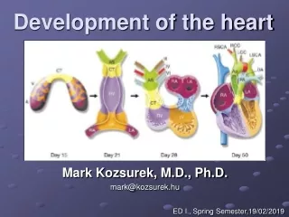
Development of the Heart. Development of primitive heart tube. It develops early in the middle of 3 rd week , from aggregation of splanchnic mesodermal cells , in cardiogenic area , ventral to pericardial coelom, and dorsal to yolk sac.

E N D
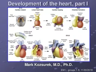
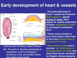
Development of primitive heart tube • It develops early in the middle of 3rdweek , from aggregation of splanchnic mesodermal cells, in cardiogenic area ,ventral to pericardial coelom, and dorsal to yolk sac. • They form 2 angioblastic cords that canalize to form 2 endocardial hearttubes. B,transverse C,longitudinal
After lateral folding ofembryo,2 endocard.tubes fuse to form…. Single hearttube (C,D) T.S of 21,22 days. • This heart tube lies inside thepericardialcavity, its dorsal wall is connected to foregut by dorsal mesocardium (D,22 days). • The central part of dorsal mesocardium degenerates ,forming transverse passage dorsal to heart ,called transverse sinusofpericardium,(E,F) schematic & T.S of 28days.
The layers of primitive heart wall : • T.S in D, at 22 days and in F at 28-days , showing : • Thin endothelial tube becomes… internal endothelial lining of the heart or endocardium. • Splanchnic mesoderm surrounding the pericardial coelom becomes….. primordial myocardium (muscular wall of heart). • Thin endothelial tube is separated from thick muscular tube (myocardium) by gelatinousC.T. (cardiac jelly)…. Forming AV septum & valves. • Visceral pericardium is derived from mesothelial cells and forms the epicardium.
After head folding of embryonic disc : • A,B,long. sections as the head fold develops (during 4th week) , heart tube & pericardial cavity lie ventral to foregut and caudal to oropharyngeal membrane. • The position of heart tube is reversed ,it lies dorsal to pericardium. • C,Long. Section, during 4th week showing : complete head folding and reversion of heart tube , pericardium & septum transverse (future central tendon of diaphragm). • Note also the heart tube lies inside the pericardial cavity.
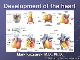
The primitive heart tubeelongates and develops alternate dilatations and constrictions :1-truncus arteriosus. 2-bulbus cordis. 3-primitive ventricle. 4-primitive atrium. 5-sinus venosus. • Truncus arteriosus is continous cranially with aortic sac ,from which aortic arches develop. • Sinus venosus has right & left hornes . • Each horn receives umbilical, vitelline ,& common cardinal veins from the chorion, yolk sac & embryo, respectively. Ventral veiw ,By the end of 4th week • Heart tube bends upon itself,giving rise an s-shaped heart,then u-shaped.
Bulbus cordis &ventriclegrow fasterthan other regions, so the heart bends upon itself,forming U-shaped bulboventricular loop (by the end of 4th week). The atrium & sinusvenosus also come to lie dorsal to truncus arteriosus, bulbus cordis & ventricle.
Blood Flow through the Primitive Heart : • By the end of 4th week,unidirectional blood flow begins at sinus venosus by peristalsis- like waves. A • Blood passes through sinuatrial valves into atrium… Atrioventricular canal … ventricle.. Bulbus cordis… Truncus arteriosus… aortic sac… aortic arches (arterial channels) … 2 dorsal aortae… into body of embryo, yolk sac , and placenta. A A,sagittal section of primordial heart(24 days),showing blood flow. B,dorsal view of heart (26 days) ,illustrating hornes of sinus venosus – Note also dorsal location ofprimordial atrium & sinus venosus.
C,ventral view of heart (35 days),Note the aortic arches arising from aortic sac and terminate in the dorsal aortae.
Partitioning of the primitive Heart • Dividing of A-V canal , primitive atrium & primitive ventricle….. Begins at the middle or end of 4th week. • It is completed by the end of 5th week. • These processes occur concurrently. A, sagittal section of primordial heart (24days),showing blood flow.
Partitioning of Atrioventricular Canal : • At the end of 4th week,2 endocardialcushions on dorsal & ventral walls of atrioventricular canal , develop from mesenchymal cells of cardiac jelly. (B) • During 5th week, the AV- endocardial cushions meet and unite in the middle line to form a septum and divide the common A-V canal into right & left A-V canals. (C,D) • Endocardial cushions also form the AV- valves + membranous septa of interventricular septum. • Note in D,cronal section ,begining of development of interatrial & intervent.septa.
Partitioning of primordial Atrium : • It begins at the end of 4th week by development of 2 septa. 1-Septum primum :a thin crescent-shaped membrane grows from the roof of common atrium into the fusing endocardial cushions dividing common primitive atrium into right & left halves. -Foramen primum is formed to pass oxyg.blood from righ to left atrium. It disapears as septum primum fuses with the endocard.cushions,(A1-C1). • Before closure of foramen primum , perforationsappear in central part ofseptum primium…coalesce to formForamen Secundum (C1-D1). • A1 to D1… coronal sections • A to D… views of interatrial septum from right side.
2-Septum secundum : a crescentic muscular memb.grows and descends from roof of atrium during 5th week. It overlaps foramen secondumin septumprimum . • The gap between the lower free border of S.secundum and the upper edge of S.primum… form ‘’foramen ovale’’. • Cranial part of S.primum disappears and remaining part of S.primum which attached to endocardial cushions… forms flaplikevalve of the foramen ovale.
In the fetus (before birth) … the pressure is higher in right atrium than in the left and highly oxygenayed blood flows directly from right atrium to leftatrium through open foramen ovale. • After birth … when the circulation of the lungs begins & the blood pressure in left atrium rises ,the upper edge of septum primum is pressed against the upper limb of septum secundum…. This will close theforamen ovale ,forming a completepartition between the 2 atria. • An oval depression in the lower part of interatrial septum of right atrium…. The fossa ovalis is a remnant of the foramen ovale.
Left side embryonic cardiovascular system (26 days) 4 –week embryo :
Changes in Sinus venosus : (A) It consists of body and 2 hornes,right & left.each horn receives 3 veins1- Vitelline vein from yolk sac. 2- Umbilical vein from placenta, 3-Common cardinal vein from body of embryo. • (B) Later , due to shuting of blood from left side to rightside in theconnection by anastomosis between the 2 anterior cardinal veins. this shunt becomes left brachiocephalic vein… (C)
Changes on left side : (B,C) 1- left horn & body of sinus venosus form the coronarysinus. 2-left common cardinal vein becomes small to form oblique vein of left atrium. 3- left vitelline & leftumbilical veins,degenerat.
Changes on right side: 1- The right horn becomes absorbed into right atrium to form its smooth part ,sinusvenarum. 2- Right common cardinal vein enlarges to form SVC. 3- Right vitelline vein becomes IVC. 4- Right umbilical vein disapears.
What happen to Sinus Venosus to share in formation of Right Atrium?1- left horn becoms the coronary sinus. 2- right horn becomes incorporated into wall of right atrium to form the smooth part (sinus venarum)… B, 8-weeks3- The remainder of the wall of right atrium + conical muscular pouch (auricle)….. have rough trabeculated area and derived fromprimordial atrium.
4-The smooth part , (sinus venarum ) & rough part (primordial atrium) are demarcated internally by a ridge, crista terminalis. -crista terminalis + valves of IVC + valves of coronary sinus are derived from right sinuatrial valve. / But left sinuatrial valve fuses withS.secundum and incorporated with it into interatrial septum.
Primordial pulmonary vein & Development of left atrium : • At first, a single common pulmonary vein is seen opening in left atrium ,just to left of S.primum. • Most smooth part of left atriumis derived fromincorporation of the singlecommon primordial pulm. veinat 5th week, (A & B). • then absorption of the 2-pulm.veins at 6th week , (C). • lastly , aborption of the 4- pulm.veins into left atrium , with separate orifices at 8th week. (D). • Left auricleis derived fromprimordial left atrium.
Embryological origin of the definitive atrium: Right Atrium Left Atrium 1-Its rough part + auricle from Right ½ of primitive atrium. 2-right ½ of A-V canal. 3- Its smooth part from Absorbed right horn of sinus venosus. 1- Its rough part +auricle from left ½ of primitive atrium. 2- left ½ of A-V canal. 3- Its smooth part from Absorbed part of pulmonary veins.
Development of muscular part of interventricular septum : • Primordial muscular interventricular( IV )septum arises in the floor of ventricle , as thick crescentic fold with concave free edge. • This septum subdivides the original ventricular cavity incompletely into right & left ventricles that communicate together through IV foramen. • This foramen closes by theend of 7th week as the 2 bulbar ridges fuse with the endocadial cushion. • A-sagittal section 5th week. • Coronal section.6th week.
Incorporation of the proximal part of bulus cordis into the ventricles • A sagittal s.at 5th w., showing the bulbus cordis in the primitive heart. • B coronal s.at 6th w. after incorporation of the proximal part of bulbus cordis into the ventricles to forms : • In right ventricle …Conus arteriosus(infundibulum), which gives origin of pulmonary trunk. • In left ventricle…. Aortic vestibule part of ventricular cavity just inferior to aorticvalve.
Closure of IV foramen & formationof membranous part of IV septum result from fusion of the following : 1-right bulbar ridge. 2-left bulbuar rige. 3-fused endocardial cushions. • A,sagittal s.at 5th w. • B, coronal s.at 6th w.after incorporation of the proximal part of bulbus cordis into the ventricles. • C,5th w.,showing the bulbar ridges & fused endocardial cushions. • D,6th w., proliferation of endocardial cushions to diminish I V foramen. • E,7th w.,fusion of bulbar ridges + extensions of endocardial cushionsupward with aortico-pulmonary septum and down with muscular I V septum to close I V foramen , so memb. IVseptum is formed
Cavitation of Ventricular Walls • Leads to formation of spongy muscular bundles (trabeculae carneae). • These bundles become the papillary muscles & tendinous cords (attached to the cusps of tricuspid & mitral valves). • A-5 weeks. • B-6weeks. • C-7weeks. • D-20 weeks.
Partitioning of distal partof the BulbusCordis & Truncus Arteriosus : • A, 5th w. ventral v.of heart. • B,5th w. transverse sections of truncus arteriosus & bulbus cordis,illustrating truncal & bulbar ridges. • C,5th w. truncal & bulbar ridges , after removal of ventral wall of heart & truncus arteriosus. • D,heart after partitioning of truncus arteriosus into aorta & pulmonary trunk. • E, transverse sections through newly formed aorta & pulm.trunk showing aortico-pulmonary septum. • F,6th w.removal of ventral wall to show aotico-pulmonary septum.
Partitioning of distal partof the BulbusCordis & Truncus Arteriosus : • G,diagram illustrating the spiral form of aortico-pulmonaryseptum. • H,drawing showing aorta & pulmonary trunk twisting around each other as they leave the heart.
Partitioning of distal partof the BulbusCordis & Truncus Arteriosus : • During 5th w. firstly , a right &left bulbar ridges are developed in the lower part. • Another ant.& post. Bulbarridges in the middle part. • Right & left truncal ridges are developed in the upper part. • Bulbar & truncal ridges are developed from proliferation of mesenchymal cells of their wall. • They are also derived from neural crest mesenchyme by passing throughthe primitive pharynx
Partitioning of distal partof the BulbusCordis & Truncus Arteriosus : • as development proceeds, the ridges fuse together following a spiral course, forming aortico-pulmonary septum which has a spiral shape at the 6th week , (as in G). • This septum divides bulbus cordid & truncus arteriosus into aorta & pulmonary trunk. • Because of spiraling of aortico-pulmonary septum, pulm.trunk twistsaround the aorta. Firstly pulm.trunk lies ant.& to right of the aorta near the ventricles, then upward,it lies post. & to left of aorta.
Development of Atrioventricular Valves • A,5thw.,showing right & left AV canals and begining of valveswellings due to proliferations of tissue (subendocardial tissue) around AV canals. • B,6th w. • C,7th w. complete development of tricuspid & mitral valves….. Note also development of compelete interventricularseptum(muscular+memb.part)
Development of aortic & pulmonary valves : • Results after development of bulbar & truncal ridges and formation of aorticopulmonary septum. • 3 Semilunar valves begin to develop from 3 swellings ofsubendocardial tissue around aortic & pulmonary orifices. • These swellings are hollowed out to form the thin walled semilunar cusps.
Development of aortic & pulmonary valves • A, long. Section showing bulbar & truncal ridges. • B, transverse section of bulbus cordis. • C,fusion of bulbar ridges. • D,formation of walls & valves of aorta & pulmonary trunk. • E, rotation of the vessels & the valves. • F, long.sections showing hollowing & thinning of valve swelling to form the cusps.
Development of conducting system : • Sinuatrial (SA) node begins to develop during 5th w.as it is present in right wallof sinus venosus. • SA-node is incorporated into wall of right atrium with sinus venosus. SA-node is located high in the rightatrium ,near entrance of SVC. • Right sinuatrial valve (cranial part)…. Forms crista terminalis,but the caudal part …forms the valves of IVC & coronary sinus.
Development of conducting system : • Left sinuatrial valve is incorporated into the interatrial septum forming AV-node & bundle ,which are located superior to endocardial cushions. • Right & left bundle branches arising from AV-bundle , pass from atrium intothe ventricular myocardium. • A band of C.T. grows in from the epicardium and separates the muscle of atria from that of ventricles to form the cardiac skeleton (fibrous skeleton of heart).
Atrial Septal defects (ASD) • There are 4 types of clinically significant types of ASD :1-ostium secundum defect. (with patent oval foramen). 2-endocardial cushion defect. (with ostium primum defect). 3-sinus venosus defect. 4-common atrium…. Rare cardiacdefect ,inwhich the interatrial septum is absent due to failure of septum primum & septum secundum to develop.
Atrial septal defect (ASD):(ostium secundum defect)A probe patent oval foramen : • A,normal postnatal, right veiw of interatrial septum after adhesion of septum primum toseptum secondum. • A1,interatrial septum, illustrating development of oval fossa in right atrium. • B and B1,note incomplete adhesion of septum primum TO septum secundum and development of a probepatent oval foramen.
Various Types of Atrial Septal Defect (ASD) in the right aspect of interatrial septum : The most common form of ASD is patent oval foramen: • A, patent oval foramen due to abnormal resorption orperforations of septum primum, (in abnormal locations), during formation of foramen secondum. • B, patent oval foramen due to excessive resorption of septumprimum ‘’short flap defect’’.
Various Types of Atrial Septal Defect (ASD) in the right aspect of interatrial septum : Patent oval foramen : • C,patent oval foramen ,resulting from an abnormallylarge oval foramen because of defective development ofseptum secundum ,so a normal septum primum will not close the abnormal oval foramen at birth. • D, patent oval foramen resulting from a combination of an abnormally large ovalforamen + excessive resorption of septum primum.
Various Types of Atrial Septal Defect (ASD) in the right aspect of interatrial septum : • E, a deficiency of fusion of endocardial cushions with septum primum and AV septal defect results and leads to a patent foramen primum -Ostium primum defect…. Less common. • F,sinus venosus ASDs(high ASDs) in the superior part of interatrial septum close to entry of SVC…. Rare type,results from incomplete absorption of sinus venosusinto right atrium and/or abnormal development of septum secundum.
Tetralogy of Fallot : • It contains 4 cardiac defects : 1- Pulmonary stenosis(obstruction of right ventricular outflow). 2- Ventricular Septal Defect (VSD). 3- Dextroposition of aorta(overriding aorta). 4- Right ventricular hypertrophy. • cyanosis is one of the obvious signs of tetralogy .
Ventricular Septal Defects (VSDs):Membranous VSD …. Is the most common type. • Results from incomplete closure of IV foramen due to failure of development of memb. part of IV septum. • Large VSDs with excessive pulmonary blood flow & pulm.hypertension result in dyspnea (difficult breathing) + heart failure.
Muscular VSD : • Due to excessive cavitation ofthe muscular part of the interventricular septum….. Producing multiple small defects (Swiss Cheese VSD). • Or absence of the IV septum--Single ventricle + Transposition of aorta & pulmonary trunk. • Complication: heart failure and death. • This diagram showing transposition of great arteries (TGA) which leads to cyanosis.VSD+ASD allow mixing arterial & venous blood. • Transposition results from that the aortico-pulmonary septum descends straight (instead of spiral).
The Aortic Arches Derivatives : • During the 4th week, as the pharyngeal arches develop, they are supplied by the aorticarches. • Aortic arches arise from the aortic sac and terminate in the dorsal aorta. • There are 6 pairs of aorticarches, but they are never present at the same time. • During 8th w.,the primitive aortic arch pattern is transformed into final fetalarteries.
Left side embryonic cardiovascular system (26 days) 4 –week embryo : • The paired dorsal aortae fuse to form a single dorsal aorta, just caudal to the pharyngeal arches. • Branches of the dorsal aorta : 1- Cervical dorsal intersegmental arteies join to form vertebral artery on each side (7th cervical intersegmental artery forms the subclavian artery). 2- Thoracic dorsal intersegmental arteries persist as intercostal arteries.3- in the lumbar region, they persist forming lumbar arteries, but 5th lumbar enlarge and forms common iliac artery.4- in the sacral region, they form lateral sacral arteries , but the caudal end of dorsal aorta becomes the median sacral artery.
The aortic Arches : • A, left sided-embryo (26-days) showing the pharyngeal arches. • B, schematic drawing showing left aortic arches arising from the aortic sac. • C, an embryo (37days), showing the single dorsal aorta and degeneration of most of the first two pairs of aortic arches.
Development of the final fetal arterial pattern : • A, aortic arches at 6 weeks, note largely disappearance of the first two pairs of aortic arches. • B,aortic arches at 7 weeks, showing normal degeneration of aortic arches and dorsal aortae. • C, final arterial arrangement at 8 weeks, note open ductus arteriosus. • D, 6-month-old infant, note the final arrangement of the vessels - and that the ascending aorta & pulmonary arteries are smaller in C than in D. Note also, obliterated & fibrosed ductusarteriosus forming … ligamentum arteriosumwithin few days after birth.