Download
1 / 15
210 likes | 1.27k Views
Lower Back Pain Therapy. Christine Mai, MD Department of Anesthesiology Boston University Medical Center. Lower Back Pain. One of the most common problem seen by pain specialists Second to headaches as cause of chronic pain Major cause of work disability worldwide

E N D
Lower Back Pain Therapy Christine Mai, MD Department of Anesthesiology Boston University Medical Center
Lower Back Pain • One of the most common problem seen by pain specialists • Second to headaches as cause of chronic pain • Major cause of work disability worldwide • Multifactorial Causes: congenital, traumatic, degenerative, myofascial syndrome, inflammatory, infectious, metabolic, psychologic, cancerous, or referred pain from retroperitoneal disease processes
Anatomy of Lumbar Spine • Anterior components: • Vertebral bodies • Intervertebral disks • Anterior/Posterior longitudinal ligaments • Posterior components: • 2 pedicles • 2 transverse processes • 2 lamina • Spinous process • Innervation: • Sinuvertebral branches arises before spinal nerve divides into anterior and posterior rami, innervates posterior longitudinal ligament, posterior annulus fibrosis, periosteum, dura and epidural vessels • Posterior Rami innervates paraspinal structures
Lumbar Facet Joint • Paired facet joints connect vertebrae in the spine • Important for both range of motion and stability • Painful when become arthritic • Facet arthropathy can further cause back spasm and referred pain frequently indistinguishable from sciatica or discogenic radicular pain • Each facet joint is innervated by medial branches of posterior primary rami, above and below the joint • Medial branch crosses upper border of the lower transverse process in groove between root of transverse process and superior articular process
Lumbar Medial Branch Facet Injection • Performed under fluoroscopy with patient in prone position • Views: AP and 30o oblique (Scotty dog view) • Insert a 22 gauge spinal needle 5-6cm lateral to spinous process, directed medially to upper border of root of transverse process • Insert at three levels (ie. L3-4, L4-5, L5-S1) • Medication: 40-80mg Triamcinolone or Methylprednisone and local anesthetic or perservative free NS 30 degree oblique view with needles in "eye of Scotty dog"
Radiofrequency Themocoagulation (RFTC) • Ablates nerve branches utilizing heat current flows from active electrode incorporated in special needle • Temperature 60-90oC for 1-3mins to ablate nerve without excessive tissue damage • Performed under fluoroscopy-important to be exactly within “eye of Scottie dog” • Electrical stimulation (2 Hz for motor response, 50 Hz for sensory response) via electrode and impedence measure help confirm correct position • Prolongs pain relief for 3-12 months • Utilized for medial branch facet rhizotomy, trigeminal rhizotomy, dorsal root rhizotomy, lumbar sympathetomy
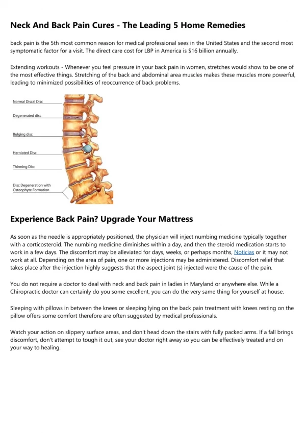
Lumbar Radiculopathy • Lower back pain radiating down lower extremities • Sensations of pain, paresthesia, numbness • Associated with herniated disks, DJD, nerve impingement • Sciatica-compression of lower nerve roots producing pain along sciatic nerve • Paracentral compression of cauda equina in dural sac can cause bilateral LLE pain, urinary retension, fecal incontinence • Inflammation results from nerve root compression MRI Lumbar Spine Herniated disks
Epidural Steroid Injections • Effective pain relief for radiculopathy • Relieves inflammation, edema and irritation by reducing swelling of nerve root, blocking c-fibers, stabilize nerve membranes, and decrease ectopic discharge from inflamed tissue • Inflammatory mediators leak into epidural space rather than subarachnoid space
Translaminar Lumbar Epidural Steroid Injection • Performed under fluroscopic guidance • Views: AP and Lateral • Maybe be left or right differentiated depending on patient’s pain location • 20 gauge Touhy needle • Loss of resistance to air/saline technique • Medications: Triamcinolone 40-80mg or Methylprednisolone 40-80mg injected with local anesthetic or with perservative free NS • Local anesthetic provides immediate pain relief until steroid inflammatory response takes place in 12-48hr Epidural space highlighted by red arrows
Transforaminal Lumbar Epidural Steroid Injection Spread of steroid injection Needle entering transforaminal epidural space • Performed when there is correlating lesion on MRI with radiculopathy • Alternative approach to epidural space when translaminar ESI fails to give relief • 22 G spinal needle directed under fluoroscopy into foramen of affect nerve root and contrast is injected to confirm entry into epidural space prior to steroid injection • Less volume of steroid/local anesthetic needed
Risks of ESI • Bleeding • Infection • Localized tenderness • Post-dural puncture headache • Paresthesia • Anxiety-related sx: lightheadedness, nausea
Selective Nerve Root Block • Performed when there is correlating lesion on MRI with radiculopathy • Used interchangeably with transforaminal epidural steroid injection • 22 gauge spinal needle inserted under fluoroscopy lateral to spinous process, directed medially to lower border of root of transverse process • Contrast is injected to confirm injection proximal to nerve root
Lumbar Facet Injection Can you visualize the Scotty Dog? At what levels are the needles inserted?
Lumbar ESI At what level is the needle inserted? Where will the patient get the most pain relief?
Selective Nerve Root Block Which nerve roots are being blocked?