Download
1 / 27
270 likes | 439 Views
Image-transfer properties of a microCT system based on a flat panel detector. Arnulfo Martínez-Dávalos Instituto de Física, UNAM arnulfo@fisica.unam.mx. Sistema Bimodal de Imágenes (SIBI). Scintillators coupled to PS-PMTs. microCT. X-ray tube. Flat panel detector. microPET. Aims.

E N D
Image-transfer properties of a microCT system based on a flat panel detector Arnulfo Martínez-Dávalos Instituto de Física, UNAM arnulfo@fisica.unam.mx
Sistema Bimodal de Imágenes (SIBI) Scintillators coupled to PS-PMTs microCT X-ray tube Flat panel detector microPET
Aims • To develop radiological imaging systems for small rodents using state of the art technologies • To integrate different imaging techniques in order to provide both anatomical and functional information • To provide advanced training in all related areas (radiation detection, nuclear techniques, imaging, preclinical studies, etc.) • To collaborate in basic biomedical research using in vivo molecular imaging techniques
Why study mice? • Biological model par excellence • Genetically modified strains • Animal models of neoplasic or neurological diseases • Non invasive in-vivo studies • Neurochemistry • Disease development • Longitudinal studies
The dose problem in microCT • In-vivo imaging allows longitudinal studies, but … • only meaningful if the subject is not altered! • Excessive dose can alter physiological functions: • 200 mGy in-vivo elevates gene expression level [Amundson et al. 2001] • 20-50 mGy in-vitro induces stress-response gene expression [Fornace et al. 2002]
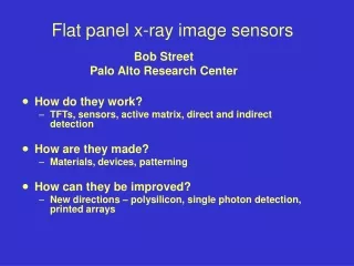
Flat panel detector • Rad-icon Shad-o-Snap 4k • 8 CMOS photodiode panels • Gd2O2S scintillator screen (Kodak Lanex fine, ~80 µm thickness) • 2000×2048, 48 µm2 pixels • 96.1 mm h × 98.6 mm w • 12 bits per pixel • 4000:1 dynamic range • USB data interface • 540 ms readout period Rad-icon Imaging Corp · 3193 Belick Street, Unit 1 · Santa Clara, CA 95054
Basic setup X-ray tube Detector X-q stage Olinca Galván, MSc Thesis, Graduate Program in Medical Physics, UNAM, 2008
mCT control panel LabVIEW v8.5, modular VI library.
Methods • X-ray spectra: Amptek CZT detector • FPN (flat field correction) • MTF : Slanted edge technique • NPS : 2D FT of relative noise distribution • DQE : MTF2 / (X Fx NPS) • Dose performance : TLD & MC • CT rec. : Feldkamp
X-ray spectra F(E,V)=a(E)kV3+b(E)kV2+c(E)kV+d(E) • Spectra • Angular distributions • Backscatter factors • Output factors • Half-value layers • Parameterization Ulises Moya, MSc thesis, Graduate Program in Medical Physics, UNAM, 2008
x : dead pixels aprox. 0.1% o : outliers aprox. 1.0% Fixed Pattern Noise
Flat field correction Non-uniform response Corrected
Linearity of response Oxford Instruments Apogee W, 1 mm Al, 1 mAs
Noise Oxford Instruments Apogee W, 1 mm Al, 1 mAs
Presampling MTF Spatial resolution at 10% MTF ~ 10 lp/mm
NPS W 40 kVp, 0.5 mm Al, 30 mR
DQE DQE = MTF2 / (X Fx NPS)
Dose measurements & simulations Carla Montaño, MSc thesis, Graduate Program in Medical Physics, UNAM, 2007
Calibration in HU Orlando Soberanis, MSc Thesis, Graduate Program in Medical Physics, UNAM, 2008
Mexican Iguana W target, 1.0 mm Al, 30 kVp, 0.5 mAs Body length ~ 4 cm
W 50 kVp, 0.5 mAs, 1 mm Al, 360º orbit, 1º steps Feldkamp, Hann filter, 0.7 cutoff frequency CT reconstruction, axial slices
Conclusions • We have built and characterized a benchtop microCT prototype • Detector performance: • Excellent spatial resolution (~10 lp/mm @ 10% MTF) • Good noise performance • Very good DQE (40-50% at low spatial frequencies) • Successful image acquisitions and tomographic reconstructions To do: • New detector, faster data transfer • Use of contrast media and/or dual energy techniques • Integration of microCT with microPET
Participants Faculty Students
Human vs. mouse Human – 70 kg Mouse – 30 g CT • 100-120 kVp • Spatial resolution ~0.4 mm • Ring geometry • Dose ~2-20 mGy microCT • 30-80 kVp • Spatial resolution ~50 μm • Flat panel detectors • Dose ~30-300 mGy PET • Spatial resolution 2-3 mm • Activity ~ 10-15 mCi • Sensitivity ~ 2-4% microPET • Spatial resolution 1-2 mm • Activity ~ 0.5 mCi • Sensitivity ~ 3.5%