Download
1 / 66
660 likes | 1.03k Views
LPN-C. Units Six & Seven Central Intravenous Therapy Pharmacology related to IV Administration. Unit Six Central Intravenous Therapy. Central Venous Access. 5 million central venous access devices (CVADs) are placed each year Increasing as the population ages

E N D
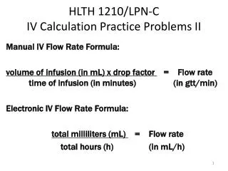
LPN-C Units Six & Seven Central Intravenous Therapy Pharmacology related to IV Administration
Unit Six Central Intravenous Therapy
Central Venous Access • 5 million central venous access devices (CVADs) are placed each year • Increasing as the population ages • Uses for central venous routes • Fluids • Medications • Blood/blood products • Chemotherapy • Nutrition • Blood samples (frequent lab tests) • Cardiac/pulmonary function assessment • Can be used repeatedly and for long periods of time
Risks/Benefits of CVAD Placement • Indications for placement of a central venous catheter (CVC) • Inadequate peripheral vascular access • Need for frequent vascular access • Hypertonic/hyperosmolar infusions • Infusion of irritating or vesicant drugs • Rapid absorption and blood/tissue perfusion • Long-term IV therapy • Contraindications for placement of a CVC • Altered skin integrity • Anomalies of the central vasculature • Cancer at the base of the neck • Cancer at the apex of the lung
Risks/Benefits (cont’d) • Contraindications (cont’d) – • Immunosuppression, septicemia • Problems with coagulation • Clavicle fracture • Hyperinflated lungs • Radiation to the insertion site area • Superior vena cava syndrome • History of venous access device complications • Main types of CVADs • Centrally inserted catheters • Peripherally inserted catheters • Centrally implanted ports • Peripherally inserted ports
Nontunneled Catheters *Single or multilumen nontunneled CVCs can be silicone or polyurethane • Inserted into the venous system from the subclavian or jugular vein by a percutaneous stick • The tip of the catheter is then advanced into the superior vena cava • Example: a Hohn catheter • Referred to as central lines, CVCs, permanent indwelling catheters, or percutaneous central venous catheters • Catheter size ranges from 24 gauge and 3½ inches to 14 gauge and 12 inches
Tunneled Catheters *Single or multilumen central venous tunneled catheters (CVTCs) are usually made of soft silicon • Dacron cuff near exit site anchors catheter in place, acts a securing device, and serves as a microbial barrier • Surgically inserted via percutaneous cutdown under local or general anesthesia • Catheter tip placed in the superior vena cava, while the other end is tunneled subcutaneously to an incisional exit site on the trunk of the body • Insertion and removal performed by doctor
Tunneled Catheters (cont’d) • Left in place for indefinite period of time • Examples are the Broviac, Hickman, and Groshong • The Groshong catheter allows fluids to flow in or out, but stays closed when not in use • Reduces need for clamping • No need for heparin flushing • NS flush every 7 days when not in use • Also available in nontunneled form
Central Catheter Insertion Sites • Subclavian vein • Accessible • High-flow • Secures easily • Less likelihood of movement of the catheter tip • Lower risk of infection • Potential complications – • Pneumothorax due to close proximity to lung apex • Laceration of the subclavian artery • Difficult to control bleeding because this is a noncompressible vessel • Internal jugular vein • Large vessel • Easily accessible
Insertion Sites (cont’d) • Internal jugular vein (cont’d) – • Short, straight pathway to superior vena cava • Potential complications – • Laceration due to close proximity to carotid artery • Difficult to immobilize and secure due to location • Greater risk of infection due to proximity to oropharyngeal secretions • Highest infection rate of all insertion sites • External jugular vein • Easy to see and locate, but not often used • Difficult to cannulate due to its valves • Tends to roll excessively *Use of the right side preferable (superior vena cava easier to access than on the left)
Insertion Sites (cont’d) • Femoral vein • If no access available in the upper body, a CVAD can be inserted into the femoral vein • Threaded into the inferior vena cava • Only done in emergencies due to increased risk of thrombosis, phlebitis, and infection *PICC lines are peripherally inserted central catheters that are placed in the basilic vein, cephalic vein, or median cubital vein • Basilic vein • Preferred choice of vessel for the insertion of a PICC line • Large in diameter • Straighter path to the superior vena cava
Central Catheter Positioning • Catheter tip should terminate in the superior vena cava • Catheter tip must never rest within the right atrium • Could traverse the sinoatrial (SA) node • Dysrhythmia • May become trapped in the tricuspid valve • Permanent damage of the valve • Requires valve replacement • Proper positioning in the superior vena cava provides – • Optimum dilution of infusates • Large volume infusate administration • Rapid administration when needed
CVC Dressing Change/Site Care • Dressing materials are to be sterile gauze or a sterile, transparent, semipermeable dressing • Dressing change is to occur at least once per week (every 7 days) • Replace dressing if damp, loosened, or visibly soiled • Clean the skin, disinfect with 2% chlorhexidine, and allow to air dry • For a PICC dressing change, replace the dressing in the same manner/position each time for evaluation purposes
Dressing Changes (cont’d) • Dressing change for a PICC (cont’d) – • If a change in position of the PICC line of more than 1 to 2 cm since insertion is noted, a chest x-ray may be indicated
Flushing/Irrigating a CVC • Maintains patency and prevents occlusion of a central line • Excessive pressure can damage the line • Never force an irrigant into the vessel • May result in embolism if thrombus is present • Never use a syringe with a barrel capacity of less than 10mL • Smaller syringes generate more pressure than larger ones • Flush with at least 10mL NS whenever the line is irrigated • Use push-pause flushing method to remove particles that adhere to the catheter lumen
PICC Lines *Single or multilumen peripherally inserted central catheters can be placed by an RN, depending on institutional policy and procedure guidelines • Usual dwelling time is 1-12 weeks, but can stay much longer • Made of silicone or polyurethane, and range in length from 33 to 60 cm • Decreases the risk of air embolism and prevents the need for frequent venipuncture • Preserves peripheral vasculature • Appropriate for home IV therapy
Midline Catheters (MLC) *An MLC is any percutaneously inserted IV line that is placed between the antecubital fossa and the head of the clavicle, and then advanced into the larger vessels below the axilla • Dwelling time is 1 to 6 weeks • Can deliver most infusates except caustic drugs and TPN that need the dilution capabilities of the superior vena cava • Verification of tip placement is per agency protocol, and placement can be completed by the RN • Useful for home IV therapy
MLC (cont’d) • Like PICC lines, placement is in the basilic vein, cephalic vein, or median cubital vein, with the basilic vein being the vessel of choice due to its size and straighter path • Line maintenance and dressing changes are the same as for PICC lines
Implantable Subcutaneous Vascular Access Devices *Also referred to as an implantable port or vascular access port (VAP) • Surgically inserted in a subcutaneous pocket under the skin without any portion of the system exiting the body • Single or double injection port connected to a catheter that is positioned in the superior vena cava • Port access must be with a noncoring needle, such as a Huber point needle or the Port-a-Cath Gripper needle, to avoid damaging the system
Implantable Ports (cont’d) • Used for long-term infusion therapy • Should not be accessed more frequently than every 1 to 3 weeks • Reduced risk of infection • Eliminates need for exit site care/dressing changes or regular flushing if not in use • Contraindicated in patients with septicemia or bacteremia • Placement is a surgical procedure • Patient will have a dressing over the incisional wound until it heals • Maintenance prior to healing is as with any surgical wound
Implantable Ports (cont’d) • A potential complication specific to a port is termed Twiddler’s Syndrome • Caused by rubbing or manipulating the skin over the implanted port • Some clients develop the habit of “twiddling” their ports, which may cause the internal catheter that is attached to the port to dislodge • Requires surgical removal and replacement of the VAP
CVC Complications • Air embolism = the entry of air into the circulatory system • May occur during insertion or removal, tubing change, or due to catheter damage/breakage • Fatality results from rapid rate of injection • Average lethal dose is 70-150cc of air, but as little as 10cc can be fatal to a gravely ill person • Signs and symptoms • Chest pain, tachycardia, thready pulse • Confusion, light-headedness, unresponsiveness • Dyspnea, pallor, hypotension • Interventions • Left-sided Trendelenburg • Clamp catheter • Administer oxygen
CVC Complications (cont’d) • Air embolism (cont’d) – • Interventions (cont’d) – • Notify the physician • Monitor vital signs • Arterial laceration = inadvertent puncture of an artery by the insertion needle or guide wire during insertion • Signs and symptoms • Hematoma, hemothorax • Hypotension, tachycardia, respiratory distress • Tracheal compression, loss of consciousness • Interventions • Apply pressure • Monitor vital signs
CVC Complications (cont’d) • Cardiac tamponade = perforation of the pericardium by the CVC that results in compression of the heart due to leakage of blood or infusates into the pericardial sac • Signs and symptoms • Cardiovascular collapse • Hypotension • Neck vein distension • Muffled heart sounds (due to fluid surrounding the heart) • Interventions • Monitor vital signs • Support the patient • Emergency resuscitation may be necessary • Aspiration of the pericardial sac
CVC Complications (cont’d) • Catheter embolism = breakage of a portion of the CVC • Due to improper insertion technique or improper administration of infusates (i.e. excessive pressure) • Signs and symptoms are dependent on where the severed portion of the CVC lodges and blocks circulation • Cardiac arrest, chest pain, hypotension, cyanosis • Dyspnea, respiratory arrest, loss of consciousness • Interventions • Institution of emergency measures • Maintain patient on bed rest • Monitor vital signs • X-ray and surgery to remove embolism
CVC Complications (cont’d) • Pneumothorax = air accumulation in the pleural cavity due to perforation of the visceral pleura during CVC insertion • Usually due to improper patient positioning • Signs and symptoms • Absent breath sounds, tachypnea • Sudden chest pain, distended chest unilaterally • Dyspnea with gasping respirations, pallor, cyanosis • Hypotension, mediastinal shift, tympanic resonance • Interventions • Administer oxygen, deep breathing and coughing • Assess breath sounds, chest expansion, vital signs • Insert chest tube • Maintain hydration and nutrition, ROM, provide rest • Semi-Fowler’s position to ease breathing
CVC Complications (cont’d) • Hemothorax = blood accumulation in the pleural cavity • Due to vessel laceration or perforation during CVC insertion • Signs and symptoms • Chest pain, cyanosis with dusky pallor • Decreased/absent breath sounds, dyspnea • Hemoptysis • Reduced hemoglobin due to blood pooling • Interventions • Administer oxygen, monitor vital signs • Remove catheter • Insert chest tube • Position to ease breathing • Provide frequent mouth care
CVC Complications (cont’d) • Hydrothorax = fluid accumulation in the thoracic cavity • Caused by vessel laceration or perforation during CVC insertion • Signs and symptoms • Chest pain, cyanosis, dyspnea • Flat, dull sound over fluid • Murmur over fluid • Vesicular breath sound absence • Interventions • Administer oxygen, monitor vital signs • Remove catheter • Insert chest tube • Aspirate pleural space fluid • Position for breathing comfort
Grading Scale for the Severity of Mechanical Phlebitis R/T PICC Line
Occluded Central Lines • Clot formation at the lumen exit • Obstruction by drug precipitates or lipid deposition • Catheter displacement • Restriction of catheter flow by sutures that have tightened around the circumference of the catheter • Coiling, kinking, or pinching of the catheter between the clavicle and the first rib • Catheter damage or transection from the repeated pressure of the clavicle and the first rib on the catheter during normal movement
Occluded Central Lines (cont’d) • Catheter pinch-off = the anatomic compression of a VAD between the clavicle and the first rib • Movement of the arm and shoulder narrows the costoclavicular space, resulting in intermittent occlusion • Diagnosis is via chest x-ray and/or the inability to aspirate blood or administer infusates unless patient’s position is changed • May result in catheter fracture • Extravasation of vesicants • Dysrhythmias • Thromboembolic formation • Catheter fragment embolization
Unit Seven Pharmacology R/T IV Administration
Pharmacokinetic Concepts • Absorption = the passage of a drug through a body surface into the tissues and the bloodstream • Distribution = process whereby a drug is transported to its intended site • Biotransformation = the metabolism of a drug within the body • Excretion = the process of removing substances from the body • Kidneys* • Lungs • Sweat glands • Gastrointestinal system
Drug Action • Plasma concentration and plasma half-life are measured in hourly intervals • Plasma concentration = the time it takes for a drug to reach its peak plasma level • When given at constant intervals over a period of time, plasma concentration reaches uniform level; will not deviate until discontinued or changes are made to administration schedule • When given at scheduled intervals, peak plasma concentration occurs immediately following administration; minimum plasma level (trough) occurs just before next dose administered
Drug Action (cont’d) • A peak blood sample is drawn immediately following the administration of a drug • A trough sample is drawn immediately before the next dose is given • Half-life refers to the time it takes the body to metabolize and eliminate ½ the original concentration of an administered drug • Abbreviated t½ • If the half-life of a drug is 4 hours, the rate at which the concentration diminishes in the body is 100% = initial bolus, 50% = 4 hours, 25% = 8 hours, 12.5% = 12 hours, 6.25% = 16 hours, and so forth
Drug Action (cont’d) *Factors affecting the action of a drug include – • Gender • Age • Body size • Occupational exposure • Lifestyle (diet, stress, exercise) • Substance use • Illness, fever • Infectious disease • Immunological disease • Tissue injury
Non-Approved Infusates for LPN-C • Blood and blood products • Antineoplastic agents • Oxytocics • Pitocin • Ergotrate • Methergine • Syntocinon • Antiarrhythmics • Hyperalimentation • TPN • PPN • Lipids
Risks of IV Drug Administration *Medication/Fluid Incompatibility = chemical or physical reaction that occurs among two or more drugs, or between a drug and the delivery device • Physical incompatibility • Cloudiness, haziness • Gas bubbles • Visible precipitation, clogging • Color changes in the tubing or filter • Chemical incompatibility • Reaction between acidic and alkaline drugs or solutions • pH instability
Risks of IV Drug Administration • Therapeutic incompatibility • Undesirable effect occurring in a patient as a result of two or more drugs being given concurrently • Produces an increase or decrease in therapeutic response • Potential complications of IV therapy • Infiltration, extravasation • Infection, phlebitis • Thrombosis, embolism • Speed shock • Allergic reaction, anaphylaxis • Pulmonary edema • Septicemia Refer to Unit 5 PPT
Drug Administration via PCA Pump *Patient-controlled analgesia (PCA) = drug administration system that enables the patient to self-administer and regulate the delivery of medication for pain control on a PRN basis • Less medication usually required by the patient due to maintenance control • Can deliver medication by IV, epidural, or subcutaneous routes • Programmed to regulate drug dosage, time intervals between boluses, and lock-out intervals (i.e. the period of time when the pump will not release medication)
INS Standards for IV Therapy *Five rights of medication administration • Right patient • Right medication • Right dose • Right route • Right time *Three checks of medication administration • Read the label of the medication as it is removed from the shelf, unit dose cart, refrigerator, or dispensing system • Read the label of the medication when comparing it with the MAR
INS Standards (cont’d) *Three checks (cont’d) – • Read the medication label again before administering the medication to the patient *Professional certification for infusion nurses • Advanced Practice Registered Nurse (APRN) • Registered Nurse (RN) • Licensed Practical Nurse – Certified (LPN-C) *Organizations providing infusion therapy must include infusion practices and standards within their policy and procedure guidelines
Basic Principles of IV Administration The nurse performing IV therapy should: • Know venous anatomy and physiology • Appropriate vein selection • Use infusion equipment appropriately • Clarify unclear orders • Refuse to follow orders that are not within the scope of safe nursing practice • Know indications, side effects, and special considerations for IV medications • Administer medications and/or infusions at the proper rate and within the ordered intervals • Use proper IV care and maintenance • Provide proper patient education
Basic Principles (cont’d) The nurse should (cont’d) – • Assess the patient’s condition and monitor the IV site for complications • Notify the physician promptly of IV complications • Know and give appropriate treatments for complications • Document all aspects of IV therapy, including patient education • Follow your institution’s policy and procedures • Abide by Nebraska’s Nurse Practice Act and the Infusion Nurses Society’s Standards of IV Practice • Keep current in research related to IV therapy
Role of the LPN-C in IV Therapy *State of Nebraska (Title 172, Chapter 102) • Perform limited IV therapy interventions under the direction of an RN or licensed practitioner. • Observe, initiate, monitor, discontinue, maintain, regulate, adjust, document, assess, plan, intervene, and evaluate with regard to IV treatment. • Provide IV interventions only when there is a licensed practitioner or RN assessing the patient at least once every 24 hours (or more frequently with significant change in therapy or condition).