Download
1 / 17
230 likes | 1.09k Views
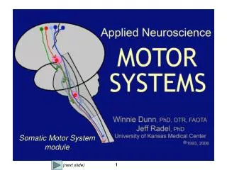
Motor system I: spinal cord circuits and motor output. Overview of the motor system Topographic relationship between spinal motor neurons and muscles Motor unit and generation of muscle force Spinal Reflexes. Overview of Motor System. Planning, initiation of voluntary movement.

E N D
Motor system I: spinal cord circuits and motor output • Overview of the motor system • Topographic relationship between spinal motor neurons and muscles • Motor unit and generation of muscle force • Spinal Reflexes
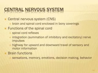
Overview of Motor System Planning, initiation of voluntary movement Sensory-motor integration, motor learning Basic movement, posture Reflex (involuntray movement)
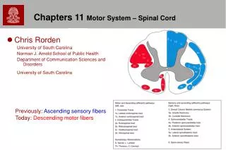
Dorsal horn Ventral horn
Motor neuron pool Motor neuron pool • In a mature animal, each muscle fiber (the muscle cell) receives axonal input from only a single motor neuron • Each motor neuron innervates multiple muscle fibers (a few to hundreds) • All motor neurons innervating a muscle (consisting of many muscle fibers) is called a motor neuron pool muscle
All motor neurons in a motor neuron pool are clustered together in spinal cord
Topographical relationship (medial-lateral) • Motor neuron pools are organized within the ventral horn, with those innervating distal muscle groups located laterally and those innervating the proximal muscles located medially
Topographical relationship (rostral-caudal) Different segments of the spinal cord innervate muscles in different parts of the body (see diagram on the right)
Motor Unit • A motor unit consists of a motor neuron and all the muscle fibers it innervates • Three types of motor units (muscles): • Fast fatigable (FF) • Powerful, but fatigue with repetitive stimulation • muscle fiber: thick, large, white (anaerobic, use glycolytic pathway to generate ATP) • Motor neuron large, fast conduction, but higher firing threshold • Slow (S) • (opposite to FF) • Muscle fiber: small, red (aerobic, use oxidative pathway to generate ATP) • Fast, fatigue-resistant (FR) • Intermediate between FF and S
Regulation of Muscle force • Progressive recruitment of motor units: S FR FF • standing (S) walking (FR) jumping (FF) • In the order of size of motor neuron • (“size principle”) - smaller motor neurons have lower threshold of firing (higher input resistance) • By changing the firing frequency • Summation of successive contractions • twitch unfused tetanus fused tetanus twitch unfused tetanus fused tetanus
Three sources of inputs to motor neurons (Output to the muscle)
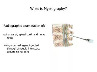
Spinal Reflex: Simple Stretch Reflex (1) Muscle stretch (activation of mechanoreceptors) (2) activation of DRG neuron (activation of group Ia afferent) { (3a) Alpha motor neuron activation contraction of the same or synergistic muscle (3b) Interneuron activation inhibition of another motor neuron reduced contraction of antagonist muscle
The dorsal root ganglion axon terminals (proprioceptor) is activated when intrafusal muscle fibers of muscle spindle detects changes in the muscle length Role of γ motor neurons: 1. Adjusting tension on intrafusal muscle (sensitivity of spindle output) 2. Fine central control of muscle contraction (“γ loop”): Descending input activates γ motor neurons in the spinal cord activation of Ia sensory afferents activation of a motor neurons innervating the homologous muscle
Stretch-induced activities in the spinal stretch reflex circuit Note that a-motor neuron has a basal firing frequency that can be modulated.
Flexion/Withdrawal Reflex Cutaneous (pain, temperature) receptors send excitation via spinal interneurons to cause: on ipsilateral side -- excitation of flexor muscle -- inhibition of extensor muscle on contralateral side -- excitation of extensor muscle -- inhibition of flexor muscle (help to maintain the balance) Both excitatory and inhibitory interneurons are involved
Three types of inhibitory interneurons in the spinal cord • 1. Group Ia inhibitory interneurons • -- coordinate opposite muscles by inhibition of motor neurons • Ranshaw cells • -- negative feedback through recurrent inhibition: branches of the motor neuron excite Ranshaw cells, which inhibit presynaptic Ia axon terminals of the same motor neuron • -- inhibit large transient excitation (prevent muscle damage) • Group Ib inhibitory interneurons • -- receiving excitation from tendon organ and cutaneous afferents to reduce motor neuron firing, to soften the touch • -- used by descending input to fine tune the muscle output