Download
1 / 43
440 likes | 691 Views
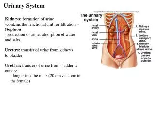
Urinary system. General outline. component. urinary organs: kidneys micturition organs: ureter, bladder and urethra. function. discharge metabolite regulate the balance of water and electrolyte endocrine function: renin, erythropoietin,

E N D
General outline • component urinary organs: kidneys micturition organs: ureter, bladder and urethra • function • discharge metabolite • regulate the balance of water and electrolyte • endocrine function: renin, erythropoietin, prostaglandin
Renal anatomic structure • Fibrosa Cortical labyrinth Medullary ray Cortex • Parenchyma Renal pyramids Renal columns Medulla
Renal histological structure Glomerulus Bowman’s capsule Renal corpuscles • nephron Renal tubules • collecting duct • juxtaglomerular apparatus
Renal cortex Renal medulla
Structure of Renal corpuscle: afferent arteriole efferent arteriole capillary network mesangial cells Glomerulus parietal layer: simple squamous Epi. Bowman’s capsule capsular cavity: filtrate visceral layer: podocytes (primary and secondary processes, slit membrane)
Glomerulus Renal corpuscle
Functions of Renal corpuscle: • Produce filtrate (primary urine) fenestrated endothelium basement membrane slit membrane of podocyte • Filtration membrane (filtration barrier) molecular weight charges • relevant factors of filtrate
Renal tubules: convoluted tubule straight tubule proximal Henle’s loops thin segment straight tubule convoluted tubule distal
Proximal tubule • Location: cortical labyrinth • LM: simple cuboidal or pyramid Epi. acidophilic cytoplasm no discrete cell margin; brush border; longitudinal striation
EM: apical canaliculi and vesicles numerous lysosomes, Mi. many lateral interdigitations microvilli on the surface membrane invaginations abundant Na+-K+ATPase
Function • reabsorb water, glucose, amino acid, protein, vitamin and inorganic salts etc. • secrete ammonia and some metabolic substances
thin segment • location: medullary ray and renal pyramids • LM: simple squamous Epi.; pale cytoplasm, • EM: a few microvilli; less organelles • Function: water, and ions pass through easily
Distal tubule • location: medullay ray and renal pyramids • LM: simple cuboidal Epi; clear cell boundary; pale cytoplasm; nuclei near lumen; without brush border; have longitudinal striation
EM: a few microvilli; many membrane invagination; many mitochondria; abundant Na+-K+ATPase • Function: reabsorb Na+ and water; excrete K+; secrete NH3
Collecting tubules: • location: medullary ray and renal pyramids • components: arched collecting tubules; straight collecting tubules; papillary ducts • simple cuboidal Epi simple columnar Epi., papillary ducts line tall columnar Epi; pale staining; distinct borders; rare microvilli and Mi. • function: similar to that of distal convoluted tubules
Juxtaglomerular apparatus: • located in a triangle area at the vascular pole of the renal corpuscles • consist of juxtaglomerular cells, macular densa and extraglomerular mesangial (polar cushion) cells • function: control water and electrolyte balance; regulate blood pressure; produce erythropoietin
Juxtaglomerular cells • smooth muscle cells of the afferent arteriole transform into the epithelial cells • cytoplasm: a few myofibrils; PAS-positive granules contained renin; abundant RER, ribosomes and well developed Golgi apparatus; • function: secrete renin and erythropoietin
Macular densa • transformed from the cells of distal tubule which near the vascular pole of the renal corpuscle • the cells become taller and narrow, arranged compactly; pale cytoplasm; nuclei located at the apex • a chemical (Na+ ) sensor
Extraglomerular mesangial cells • resemble the intraglomerular mesangial cells • gap junctions between the component of the juxtaglomerular apparatus • transmit information
Features of renal blood circulation • blood flow is large • two sets of capillary network • the diameter of afferent arterioles is larger than that of efferent, so as to facilitate filtration • the vasa recta are parallel to the Henle’s loop, so aid water reabsorption and urine concentration
Micturition organs (ureter, bladder,) Epi: transitional Epi Lamina propria: L.C.T. • mucosa • muscle layer: smooth muscle • adventitia: fibrosa serosa