Download
1 / 53
540 likes | 689 Views
The Knee: Rehabilitation Techniques. Complex joint that endures great amounts of trauma due to extreme amounts of stress that are regularly applied Hinge joint w/ a rotational component Stability is due primarily to ligaments, joint capsule and muscles surrounding the joint

E N D
Complex joint that endures great amounts of trauma due to extreme amounts of stress that are regularly applied • Hinge joint w/ a rotational component • Stability is due primarily to ligaments, joint capsule and muscles surrounding the joint • Designed for stability w/ weight bearing and mobility in locomotion
Assessing the Knee Joint • Determining the mechanism of injury is critical • History- Current Injury • Past history • Mechanism- what position was your body in? • Did the knee collapse? • Did you hear or feel anything? • Could you move your knee immediately after injury or was it locked? • Did swelling occur? • Where was the pain
History - Recurrent or Chronic Injury • What is your major complaint? • When did you first notice the condition? • Is there recurrent swelling? • Does the knee lock or catch? • Is there severe pain? • Grinding or grating? • Does it ever feel like giving way? • What does it feel like when ascending and descending stairs? • What past treatment have you undergone?
Observation • Walking, half squatting, going up and down stairs • Swelling, ecchymosis • Assessment of muscle symmetry/atrophy • What is the athlete’s level of function? • Does the athlete limp? • Full weight bearing? • Does athlete exhibit normal knee mechanics during function?
Palpation • Athlete should be supine or sitting at edge of table with knee flexed to 90 degrees • Should assess bony structures checking for bony deformity and/or pain • Soft tissue • Lateral ligaments • Joint line • Assess for pain and tenderness • Menisci
Special Tests for Knee Instability • Use endpoint feel to determine stability • Classification of Joint Instability • Knee laxity includes both straight and rotary instability • Translation (tibial translation) refers to the glide of tibial plateau relative to the femoral condyles • As the damage to stabilization structures increases, laxity and translation also increase • Valgus and Varus Stress Tests • Used to assess the integrity of the MCL and LCL respectively • Testing at 0 degrees incorporates capsular testing while testing at 30 degrees of flexion isolates the ligaments
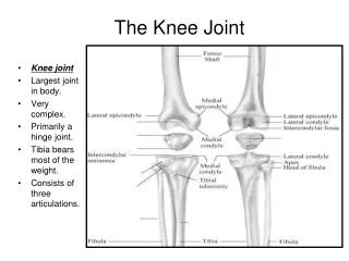
Functional Anatomy • Knee is directly affected by motions and forces occurring at the foot, ankle and lower leg • Knee transmits forces to thigh, hip, pelvis, and spine
Functional Anatomy • Considered a hinge joint: primarily knee flexion and extension • Not a true hinge joint because does have rotational and translational movements • Knee provides stability in weight bearing and mobility in movement • Joint stability dependent on ligaments, joint capsule and surrounding musculature • Unstable laterally and medially
Functional Anatomy • Collateral Ligaments • MCL: Supports medial aspect of knee • stabilizer against medial forces (valgus stress) or knee moving toward midline • Also stabilizes external rotation • LCL: supports lateral aspect of knee • Protects knee from varus (Lateral) forces or knee moving away from midline
Functional Anatomy • Cruciate Ligaments • ACL: prevents tibia from moving anteriorly • Stabilizes knee in full extension and prevents hyperextension • Also stabilizes tibia against excessive internal rotation • Secondary stabilizer from valgus and varus stress • PCL: prevents tibia from moving posteriorly.
Functional Anatomy • Menisci • Medial and lateral menisci • function to improve stability of knee • increase shock absorption • distribute weight over a larger surface area • Move posteriorly during extension and anteriorly during flexion
Functional Anatomy • Patella • Along with quadriceps and patellar tendon form the extensor mechanism • Aids knee during extension by lengthening the lever arm of the quadriceps muscle • Distributes compressive stresses on the femur by increasing contact area between patellar tendon and the femur • Protects patellar tendon from friction
Functional Anatomy • Patella • Tracking of the patellar tendon dependent on pull of quadriceps muscle, patellar tendon, depth of femoral condyles and shape of patella
Functional Anatomy • Muscle Actions • For knee to function properly numerous muscles work together in a highly complex coordinated fashion • Knee movement requires various muscles to act as agonist, antagonists, synergists, stabilizers and neutralizers • Athletic movements require multi-planar forces so rehabilitation of these muscles should mimic these movements
Rehabilitation Techniques • Phase I: • Control pain, swelling and assist tissue healing • P.R.I.C.E. • For some knee injuries may require immobilizer or rehabilitation brace • Modalities • Maintain core and cardiorespiratory function
Rehabilitation Techniques • Phase II: ROM • Early ROM can minimize harmful changes in ligaments due to immobilization • AROM: Heel slides, active hamstring stretch, squats, quad sets • PROM: Prone/supine leg extensions with or w/o assistance. Static quad, hip, groin, glut, ITB, seated calf and hamstring stretching • AAROM: Wall Slides, heel slides with contralateral limb assistance • PNF
Rehabilitation Techniques • Phase III: Strengthening. • Overload principle, but not too early or too aggressive • can harm healing tissue • Gently progressive: isometric to isotonic to isokinetic to plyometric to functional activity • Closed chain exercises proven to be more effective for knee rehabilitation • Can implement early on in most knee rehabs • OKC can increase anterior tibial shear
Rehabilitation Techniques • Isometrics: Quad Sets, Glut Sets, Hamstring Sets. Co-contraction • Isotonic: SLR-hip flex, ext. abduction and adduction. Squats; DL & SL, lunges; single, multiplane, step ups forward and lateral. Deadlifts; double leg and single leg. Clams • Proprioception: Progressive balance exercises • Can add unstable surfaces, perturbations, ball toss and/or rotation to exercises as they progress
Rehabilitation Techniques • Phase IV: Functional progressions return to sport activity • Running progression: straight ahead at ½ speed to ¾ speed to full sprint to change of direction lateral movement @ ½ speed to ¾ speed to full speed. • Sport specific movements and activity
Rehabilitation Techniques • Phase V: Maintenance and monitoring of return to sport • All phases should include core and cardiorespiratory exercises • Athlete should be comfortable and confident in their progression • Use pain and swelling as guide for progression.
Recognition and Management of Specific Injuries • Medial Collateral Ligament Sprain • Cause of Injury • Result of severe blow or outward twist – valgus force • Signs of Injury - Grade I • Little fiber tearing or stretching • Stable valgus test • Little or no joint effusion • Some joint stiffness and point tenderness on lateral aspect • Relatively normal ROM
Signs of Injury (Grade II) • Complete tear of deep capsular ligament and partial tear of superficial layer of MCL • No gross instability; slight laxity • Slight swelling • Moderate to severe joint tightness w/ decreased ROM • Pain along medial aspect of knee • Signs of Injury (Grade III) • Complete tear of supporting ligaments • Complete loss of medial stability, meniscus disruption • Minimum to moderate swelling • Immediate pain followed by ache • Loss of motion due to effusion and hamstring guarding • Positive valgus stress test
Care • RICE for at least 24 hours • Crutches if necessary • Knee immobilizer may be applied • Move from isometrics and STLR exercises to bicycle riding and isokinetic • Return to play when all areas have returned to normal • Continued bracing may be required
Care • Conservative non-operative approach for isolated grade 2 and 3 injuries • Limited immobilization (w/ a brace); progressive weight bearing for 2 weeks • Follow with 2-3 week period of protection with functional hinge brace • When normal range, strength, power, flexibility, endurance and coordination are regained athlete can return • Some additional bracing and taping may be required
Specific Injury considerations • MCL • Most commonly injured ligament • Usually treated conservatively w/o surgery • Grade 1 & 2 weight bearing as soon as tolerated. • Grade 2 may require use of protective functional knee brace for 2 weeks • Grade 3 protective rehabilitation brace for 4 to 6 weeks • Locked in 0 to 90 degrees of movement
MCL • Grade 1 • Can begin early ROM and isometric exercise 1-2 days after injury • May return to activity fairly quickly • Grade 2 • May require 4-5 days of rest to allow inflammation to subside before starting rehab ex. • 4-6 week recovery period • Exercise bike and closed chain exercises can begin for grade 1 & 2 sprains as early as it is tolerated
MCL • Grade 3 sprains • May take up to 3 months to return • Brace for 4-6 weeks, non-weight bearing for 3 weeks. • Can remove brace for treatment and rehab • Rehab limited to isometrics and straight leg exercises • After the brace is discontinued can progress rehab similar to Grade 1 & 2 sprains
Functional and Prophylactic Knee Braces • Used to prevent and reduce severity of knee injuries • Provide degree of support to unstable knee • Can be custom molded and designed to control rotational forces and tibial translation
Lateral Collateral Ligament Sprain • Cause of Injury • Result of a varus force, generally w/ the tibia internally rotated • Direct blow is rare • Signs of Injury • Pain and tenderness over LCL • Swelling and effusion around the LCL • Joint laxity w/ varus testing • Care • Following management of MCL injuries depending on severity
Anterior Cruciate Ligament Sprain • Cause of Injury • MOI – athlete decelerates with foot planted and turns in the direction of the planted foot forcing tibia into internal rotation • May be linked to inability to decelerate valgus and rotational stresses - landing strategies • Male versus female • Research is quite extensive in regards to impact of femoral notch, ACL size and laxity, malalignments (Q-angle) faulty biomechanics • Extrinsic factors may include, conditioning, skill acquisition, playing style, equipment, preparation time • Also involves damage to other structures including meniscus, capsule, MCL
ACL Prevention Programs • Focus on strength, neuromuscular control, balance • Series of different programs which address balance board training, landing strategies, plyometric training, and single leg performance • Can be implemented in rehabilitation and preventative training programs • Shoe Type • Change in football footwear has drastically reduced the incidence of knee injuries • Shoes w/ more shorter cleats does not allow foot to become fixed while still allowing for control w/ running and cutting
Signs of Injury • Experience pop w/ severe pain and disability • Rapid swelling at the joint line • Positive anterior drawer and Lachman’s • Other ACL tests may also be positive • Care • RICE; use of crutches • Arthroscopy may be necessary to determine extent of injury • Could lead to major instability in incidence of high performance • W/out surgery joint degeneration may result • Age and activity may factor into surgical option • Surgery may involve joint reconstruction w/ grafts (tendon), transplantation of external structures • Will require brief hospital stay and 3-5 weeks of a brace • Also requires 4-6 months of rehab
Surgical considerations • Young, highly athletic • Unwilling to change active lifestyle • Rotational instability or feeling of “giving out” with normal activities • Injury to other ligaments and/or menisci • Recurrent joint swelling • Failure of conservative treatment (intensive rehab program) • Surgery necessary to prevent early onset of degenerative changes within knee
4 to 12 month return to activity depending on various ACL protocols • Choice of grafts to use for ACL reconstruction may influence time frame • Also depends on surgeon, physical therapist and ATC
Posterior Cruciate Ligament Sprain • Cause of Injury • Most at risk during 90 degrees of flexion • Fall on bent knee is most common mechanism • Can also be damaged as a result of a rotational force • Signs of Injury • Feel a pop in the back of the knee • Tenderness and relatively little swelling in the popliteal fossa • Laxity w/ posterior sag test • Care • RICE • Non-operative rehab of grade I and II injuries should focus on quad strength • Surgical versus non-operative • Surgery will require 6 weeks of immobilization in extension w/ full weight bearing on crutches • ROM after 6 weeks and PRE at 4 months
Meniscus Injuries • Cause of Injury • Medial meniscus is more commonly injured due to ligamentous attachments and decreased mobility • Also more prone to disruption through torsional and valgus forces • Most common MOI is rotary force w/ knee flexed or extended while weight bearing • Signs of Injury • Diagnosis is difficult • Effusion developing over 48-72 hour period • Joint line pain and loss of motion • Intermittent locking and giving way • Pain w/ squatting
Care • Immediate care = PRICE • If the knee is not locked, but indications of a tear are present further diagnostic testing may be required • Treatment/rehabilitation should follow that of MCL injury • If locking occurs, anesthesia may be necessary to unlock the joint w/ possible arthroscopic surgery follow-up • W/ surgery all efforts are made to preserve the meniscus -- with full healing being dependent on location • Torn meniscus may be repaired using sutures
nonoperative • “wait and see” approach • May return to play when symptoms subside and progressive rehab plan is complete • Partial menisectomy • Removal of torn tissue • Full return in 2 to 4 weeks
Meniscal repair • Sutures used to repair torn meniscus • Brace locked in extension for 2 weeks • Brace locked in 0 to 90 degrees for an additional 2 to 4 weeks • Generally return to activity after 3 months • Meniscal transplant • Replacement of meniscus with allografts (cadaver meniscus) or synthetic tissue • Efficacy is inconsistent • Return to activity 9 to 12 months
Iliotibial Band Friction Syndrome (Runner’s Knee) • Cause of Injury • Repetitive/overuse conditions attributed to mal-alignment and structural asymmetries • Result of repeated knee flexion & extension • Signs of Injury • Irritation at band’s insertion • Tenderness, warmth, swelling, and redness over lateral femoral condyle • Pain with activity • Care • Correction of mal-alignments • Ice before and after activity, proper warm-up and stretching; NSAID’s • Avoidance of aggravating activities
Patellar Fracture • Cause of Injury • Direct or indirect trauma (severe pull of tendon) • Forcible contraction, falling, jumping or running • Signs of Injury • Hemorrhaging and joint effusion w/ generalized swelling • Indirect fractures may cause capsular tearing, separation of bone fragments and possible quadriceps tendon tearing • Little bone separation w/ direct injury • Management • X-ray necessary for confirmation of findings • RICE and splinting if fracture suspected • Refer and immobilize for 2-3 months
Acute Patella Subluxation or Dislocation • Cause of Injury • Deceleration w/ simultaneous cutting in opposite direction (valgus force at knee) • Quad pulls the patella out of alignment • Some athletes may be predisposed to injury • Repetitive subluxation will impose stress to medial restraints • More commonly seen in female athletes • Signs of Injury • W/ subluxation, pain and swelling, restricted ROM, palpable tenderness over adductor tubercle • Dislocations result in total loss of function • First time dislocation = assume fracture
Care • Immobilize and refer to physician for reduction • Ice around the joint • Following reduction, immobilization for at least 4 weeks w/ use of crutches • After immobilization period, horseshoe pad w/ elastic wrap should be used to support patella • Muscle rehab focusing on muscle around the knee, thigh and hip are key (STLR’s are optimal for the knee)
Chondromalacia patella • Cause • Softening and deterioration of the articular cartilage • Possible abnormal patellar tracking due to genu valgum, external tibial torsion, foot pronation, femoral anteversion, patella alta, shallow femoral groove, increased Q angle, laxity of quad tendon • Signs of Injury • Pain w/ walking, running, stairs and squatting • Possible recurrent swelling, grating sensation w/ flexion and extension • Care • Conservative measures • RICE, NSAID’s, isometrics for strengthening • Avoid aggravating activities • Surgical possibilities