Download
1 / 19
220 likes | 461 Views
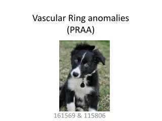
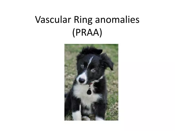
Vascular Ring anomalies (PRAA). Buster. 8 week old Border collie Regurgitation/vomiting recently since starting to feed kibble Smaller than the rest of his littermates Always hungry. Thoracic Radiographs. Cranial thoracic megaesophagus. Leftward deviation of the trachea

E N D
Buster • 8 week old Border collie • Regurgitation/vomiting recently since starting to feed kibble • Smaller than the rest of his littermates • Always hungry
Thoracic Radiographs Cranial thoracic megaesophagus
Leftward deviation of the trachea Absent left margin of the aorta
EMBRYOLOGY Vascular Ring Anomalies: Case Report and Brief Overview. Can. Vet. J. 20: 78-81(March 1979)
Embryology • The FIRST and SECOND arches regress and the internal and external carotids are formed by the dorsal and ventral aortas respectively. • The dorsal aortas between the third and fourth arches regress leaving the THIRD arch as part of the internal carotid artery. • The left FOURTH arch enlarges to form the main aortic arch while • The right FOURTH arch becomes the right subclavian artery. • The FIFTH arch is vestigial and of no apparent clinical significance. The SIXTH arches become the pulmonary arteries. • The left SIXTH arch retains its connection with the left dorsal aorta as the ductusarteriosus. • Many variations of the vascular ring anomaly based on degrees of patency and association with other cardiac lesions have been described. Vascular Ring Anomalies: Case Report and Brief Overview. Can. Vet. J. 20: 78-81(March 1979)
VRAs • Congenital malformations of the major arteries of the heart that entrap the intrathoracic esophagus causing esophageal obstruction • PRAA is the most common: R aa rather than the L 4thaa becomes the functional aorta • Causes: Circular entrapement of the esophagus • Aorta on the right, ligamentumarteriosumdorsolaterally on the left, PT on the Left and the heart base ventrally
5 MAIN FORMS OF VASCULAR RING ANOMALIES • 1. Double aortic arch: both fourth arches maintain their attachment to the adult descending aorta. • 2. Right aortic arch with left ductusarteriosus: ring formed by the normal ductusarteriosus from the left pulmonary arch joining to the abnormal right aortic arch. • 3. Left aortic arch with right ductusarteriosus: ring formed by the abnormal communication between the right pulmonary arch and the normal aortic arch. • 4. Aberrant right subclavian artery: origin of the artery such that the vessel must pass retro-esophageal and thus put pressure on the esophagus, trachea, etc. • 5. Aberrant left subclavian artery: as for the aberrant right subclavian artery.
Clinical Signs • Result from constricted esophagus • Regurgitation of solid food after weaning • Wt loss with failure to thrive despite good appetite • Moist cough, fever, dyspnea: if aspiration pnuemonia present • PE: thin stunted animal • Breed predisposition: Irish Setters and German Shephards
Diagnosis • Thoracic rads: • cranial megaesophagus cranial to the base of the heart • Leftward deviation of the trachea near the cranial border of the heart • Esophagram: Confirm location of esophageal constriction and evaluate esophageal motility to help give prognosis of function post-sx
Diagnostics • CT: to confirm type of VRA present. • Some animals may have more than 1 anomalous vessel • Will give surgeon more information to plan sx accordingly • Echocardiogram • May be able to identify anomalous vessel and/or other congenital abnormalities
PRAA DUCTUS ARTERIOSUS AORTA MEGAESOPHAGUS Left pulmonary arch
Multiple anomalous vessels • 3 month old GSD: PRAA, PDA, persistent retroeshageal left subclavian artery, persistent left cranial vena cava and left azygous vein Multiple vascular anomalies in a regurgitating German shepherd puppy. Christiansen, Snyder, Buchanan, and Holt. Journal of Small Animal Practice(2007) 48, 32-35
Treatment • Surgical ligation and transection of the ligamentumarteriosum • Tx aspiration pneumonia if present • Esophageal hypomotility and regurgitation may persist elevated feedings and promotility medications
PRAA Thrall. Textbook of Veterinary Radiology. Fifth edition; 2007. p 505-506
Double Aortic Arch Thrall. Textbook of Veterinary Radiology. Fifth edition; 2007. p 505-506
Aberrant Right Subclavian Thrall. Textbook of Veterinary Radiology. Fifth edition; 2007. p 505-506