Download
1 / 16
160 likes | 272 Views
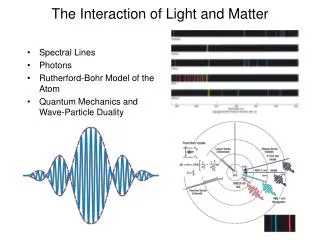
Controlling the interaction between light and matter confined in nanoscale. Leonardo de S. Menezes Departamento de Física Universidade Federal de Pernambuco 50670-901 Recife-PE, Brasil. lmenezes@df.ufpe.br www.df.ufpe.br/~lmenezes.

E N D
Controlling the interaction between light and matter confined in nanoscale Leonardo de S. Menezes Departamento de Física Universidade Federal de Pernambuco 50670-901 Recife-PE, Brasil lmenezes@df.ufpe.br www.df.ufpe.br/~lmenezes Limits and Interfaces in Sciences / Kumboldt-Kolleg São Paulo-SP, 28th - 30th October 2009
Outline of the talk Motivation Using scanning probe tech-niques (SNOM) for controlling and manipulating confined light in microresonators, as well as to control the interaction of single nanoparticles with it. • Introduction • Scanning near-field optical microscope (SNOM) probe for controlling Raman microlaseraction • SNOM probe as a tool for controlling the interaction of a nanoscopic light emitter with confined electromagnetic field
~33 m 15 µm St. Paul´s cathedral, London Lord Rayleigh, 1878 33 m 100 m Whispering gallery modes Light is trapped in a whispering gallery mode by successive total internal reflections, travel-ling in a great circle along the cavity's perimeter. Microspheres as optical cavities Represent optical resonators with ultra-high Q-factors and small mode volumes. • Easily produced by melting an optical fiber with a CO2 laser. • Diameters from 20 m to 200 m. • Q factors up to 1010. • May store photons for some s. • Comparison: tuning fork 550 Hz, same Q: oscillates for 4 days!!! • Modal volume V~3003 • Evanescent field allows the external coupling. Braginsky et al., Phys. Lett. A 137, 393 (1989); L. Collot et al., Eur. Phys. Lett. 23, 327 (1993).
Experimental setup Spectroscopy of the microspheres´ eigenmodes Typical spectrum measured by absorption and scattering
Experimental setup… Scanning Near-field Optical Microscopy • Constant distance(~10 nm) betweenthemicrospheresur-faceandthe SNOM tip via a shear force control loop. • Tip-limited(~50 nm) opticalresolution. • Allowsgetting a topogra-phicalimage. 20mm
2. Scanning optical near-field probe for controlling Raman microlaser action
4 mm =70m Q=3108 Raman microlaser =795nm For Q = 109, Pthreshold = 4.3 W world record!
Controlling ultralow threshold Raman microlasing action Laser mode (@ 814 nm) Pumpmode (@ 795 nm) Tip reduces the Q-factor of the WGM laser threshold increases A. Mazzeiet al., Appl. Phys. Lett. 89, 101105 (2006).
3. Controlled and efficient photon transfer between two single nanoemitters mediated by WGMs
Without notch filter With notch filter Controlled coupling with a single dye-doped bead Fluorescence microscope images of a single 200 nm dye-doped bead attached to a SNOM tip
200 nm Coupling of a single nanoemitter to WGMs 200 nm in diameter dye-doped bead S. Götzinger et al., Nano Lett. 6, 1151 (2006).
Confocal laser scanning micros-cope + dip coated microspheres via scope via prism Coupling of single semiconductor quantum dots: S. Götzinger et al., J. Opt. B: Quantum Semiclass. Opt. 6, 154 (2004).
Heart of the experimental setup Multimode fiber connected to PMT Prism Collimating lens Confocal microscope obejctive Monomode fiber socket Cu tube with microsphere Rotation stage Temperature stabilized Cu block Monomode fiber with collimating and focus-sing lenses Stabilized 3D piezo stack Goniometer
lexc=532nm Intensity (arb. units) =35 m Wavelength (nm) Intensity (arb. units) Wavelength (nm) Efficient photon transfer between two single nanoemitters Cavity-mediated photon transfer Our calculations show that the transfer efficiency is 106 times larger that in free space! S. Götzinger et al., Nano Lett. 6, 1151 (2006).
Conclusions We have shown how to fabricate microresonators presenting resonan-ces with ultrahigh quality factors, i.e., ultralong photon storage times. We have used silica microspheres to observe an ultralow threshold Raman microlaser action and used a SNOM probe to control it. A single nanoparticle was attached to the end of a near-field probe. The coupling to a high-Q WGM was obtained in a very controlled way. By using our setup, we have obtained cavity-mediated enhanced pho-ton transfer between two single nanoparticles. Andpretty close to ourlabs... Baía dos Porcos, Fernando de Noronha-PE Thankyou for yourattention!!!