Download
1 / 101
1.01k likes | 1.21k Views
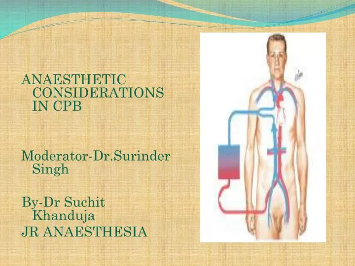
ANAESTHETIC CONSIDERATIONS IN CPB Moderator- Dr.Surinder Singh By-Dr Suchit Khanduja JR ANAESTHESIA. DEFINITION.

E N D
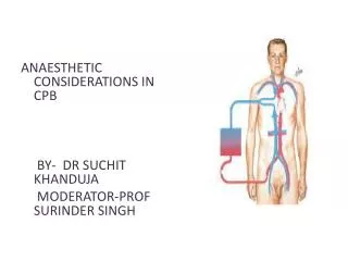
ANAESTHETIC CONSIDERATIONS IN CPB Moderator-Dr.Surinder Singh By-Dr SuchitKhanduja JR ANAESTHESIA
DEFINITION CPB is the technique whereby blood is totallyor partially diverted from the heart into a machine with the gas exchange capacity and subsequently returned to the arterial circulation at appropriate pressures & flow rates.
HISTORICAL ASPECTS • Legllois(1812) : “circulation might be taken over for short periods” • The first heart-lung machine was built by physician, John Heysham Gibbon in 1937 • Dr. Clarence Dennisled the team that conducted the first known operation involving open cardiotomy with temporary mechanical takeover of both heart and lung functions on April 5, 1951 at the University of Minnesota Hospital. • Dr.JohnGibbon(Philadelphia) 1953 : “performed ASD repair with the aid of CPB for the 1st time with the survival of patient.”
GOALS OF CPB To provide a still & Bloodless Heart with blood flow temporarily diverted to an Extracorporeal Circuit that functionally replaces the Heart & the Lung
COMPONENTS OF CPB • TOTAL CPB : Systemic venous drainage CPB Circuit External oxygenator heat exchanger External pump arterial filterSystemic circulation. • PARTIAL CPB : Portion of systemic venous return (Rt. Heart) CPB .Undiverted blood Rt. Atrium Rt. Ventricle Pul. Circulation Lt. Atrium & Lt. Ventricle Systemic Circulation.
INTEGRAL COMPONENTS OF extracorporeal circuit • PUMPS • OXYGENATOR • Heat exchanger • Arterial filter • Cardioplegia delivery system • Aortic/atrial/vena cavalcannulae • Suction/vent
PATIENT ARTERIAL LINE FILTER RESERVOIR ROLLER PUMP OXYGENATOR HEAT EXCHANGER
ROLLER PUMP • Most commonly used. • Uses Volume displacement to create forward blood flow. • Non Pulsatile Blood Flow • By compressing Plastic Tubing b/w Roller & Backing Plate
Properly set occlusion causes minimal haemolysis • Occlusion is 100% in cardioplegia &vent pumps • Each pump indepedently controlled by a rheostat • Larger tubing and lesser rotations cause minimal haemolysis
DISADVANTAGE of producing PULSATILE FLOW • Bubble Formation • Damage to Blood Components. ADVANTAGE : • Improved Tissue Perfusion • Better Preservation of Organ Function (Brain , Kidney)
CENTRIFUGAL PUMP • Series of cones that spin & propel blood forward by Centrifugal Force. • Safe • Reliable • Disposable • Simple to operate.
CENTRIFUGAL PUMP • DISADVANTAGE • Inability to generate pulsatile flow • Potential discrepancy b/w pump speed & actual flow generated. • ADVANTAGE • No back pressure when tubing is temporarily obstructed / kinked • Doesn’t produce spatulated emboli from compression of the tubing • Cannot pump large amt.of gas / gas emboli. • Less blood trauma • High vol. output with moderate pressures
Preferred over roller pumps in • Long-term CPB • In high-risk angioplasty patients • Ventricular assistance • Neonatal ECMO
Pressure-regulated pump • Operates under passive filling • After&pre-load sensitive • Pump-chamberofpolyurethane+peristalticpump • Not yet fully evaluated
OXYGENATOR • Where O2 & CO2 Exchange takes place. • Two Types : BUBBLE OXYGENATOR MEMBRANOUS OXYGENATOR
BUBBLE OXYGENATOR • Gas exchange by directly infusing the gas into a column of systemic venous blood. • A) OXYGENATING CHAMBERS:bubbles produced by ventilating gas through diffusion plate into venous blood column • Larger the No. of Bubbles ; Greater the efficiency of the oxygenator. • Larger bubbles improve removal of CO2 , diffuses 25 times more rapidly in plasma than O2 • Smaller bubbles are very efficient at oxygenation but poor in co2 removal
ADVANTAGE Easy to assemble Relatively small priming Volumes Adequate oxygenating capacity Lower cost. DISADVANTAGE Micro emboli Blood cell trauma Destruction of plasma protein due to gas interface. Excessive removal of CO2 Defoaming capacity may get exhausted with time. BUBBLE OXYGENATOR
MEMBRANOUS OXYGENATOR • Gas exchange across a thin membrane • Eliminates the need for a bubble-blood contact & need for a defoamer; so more physiological. • Blood damage is minimum • Ideal for perfusions lasting for >2-3 hours. • 2 types of membrane: • SOLID: Silicone • MICROPOROUS:polypropylene,Teflon &polyacrylamide
MEMBRANOUS OXYGENATOR ADVANTAGE • Can deliver Air-O2 mixtures. • Hemolysis • Protein desaturation • Post-op bleeding • Better platelet preservation. DISADVANTAGE • Expensive • Large priming volume • Prolonged use pores may get blocked.
CIRCUITS • Drains Venous Blood by gravity into oxygenator & returns the oxygenated blood under pressure to the systemic circulation.
VENOUS DRAINAGE • Systemic venous blood (Rt.Heart)to Oxygenator by • Direct Cannulation of SVC & IVC (BicavalCannulation) thru RA & joined to create a single drainage channel. • Single cannula into RA thru RA appendage • Mostly RA cannulation done • Bicavalcannulation done is procedure such as MVR. • Blood flow to Oxygenator (Gravity) • Height Difference B/w Venacavae & Oxygenator > 20-30 cm.
Complications • arrhythmia • bleeding • ivc/svc tear • cannulamalposition • low return • inadequate height • malposition • kink,clamp,air lock
TUBINGS IN THE CIRCUIT • Made of • PVC,Polyurethane,Silicone • I.D . Ranges from 3/16- 5/8 inches • Non thrombogenic , Chemically Inert to prevent clotting Trauma to blood elements Protein Denaturation • Smooth Internal Finish • Non Reactable Internal Surface • Durable to withstand high pressure & use of Roller pump
Disadvantages of plain circuits • Activation of platelets/coagulation factors • Post-op consumptive coagulopathyimmune reactions • More spallation Heparin coated circuits are • More hemo compatible • Cause less activation of platelets/white cells • Reduce heparin demand
INTRACARDIAC SUCTION • Blood will enter the heart Coronary venous Return Retrograde flow in AR. Bronchial Arteries CARDIOTOMY SUCTION • Spilled Heparinised Blood is Scavenged & returned back to patient. • Handheld Suckers are used to return this blood.
VENTRICULAR VENTING • Done by passing the vent from superior pulmonary vein to LA to left ventricle • Can also be done through Aortic root • Blood from LV flows to Reservoir Bag • LV Venting done to • Keep the operative field clear • Maintain Low LA & Pul.Venous Pressure • Remove air from Cardiac Chamber. • Blood from LV Reservoir Bag
RESERVOIR BAG • Collects the blood from venous drainage and cardiotomy suction passively • Blood reservoirs may be collapsible plastic bags or clear plastic hard-shelled containers. • Hard-shelled reservoirs include an integral filtration mechanism with a screen and depth filters through which blood must pass before leaving the outlet of the vessel. • Volume in the bag should not be allowed to empty to prevent massive emboli.
ARTERIAL RETURN • Ascending Aorta just proximal to Innominate Artery. • Femoral Artery in • Dissecting Aortic Aneurysm • For Reoperation • Emergency • Problems of Femoral Cannulation : • Sepsis • Formation of False Aneurysm • Development of Lymphatic Fistula. Axillary artery cannualtion done in surgeries involving aortic arch
ARTERIAL CANNULA • Is the Narrowest part of the circuit. • Should be as Short as possible. • As Large as the diameter of vessel permits.
Complications • Difficult Cannulation • Intramural Placement • Air embolism • Dislodgement of Cannula • Dissection • Arch Vessel Cannulation • Back Wall Injury
MICROPORE FILTERS: • Remove Particulate Matter (Bone , Tissue , Fat , Blood Clots etc.) • Pore Size : 30 – 40 • ULTRAFILTRATION : • Remove the excess fluid from the CPB.
PRIME FLUID • Ideally close to ECF. • Whole Blood NOT used : • Homologous Blood Syndrome. • Post Perfusion Bleeding Diathesis • Incompatibility Reactions. • Demand on Blood Banks. • Addition of Priming Fluid HEMODILUTION.
COMPOSITION OF PRIME : • Balanced salt soln. RL 1250 ml • Osmoticallyactive agent 100 ml (Mannitol, Dextran 40 , Hexastarch) • NaHCO3 50ml • KCl10ml • Heparin 1ml
PRIMING • Heme, nonheme • Decreases viscosity so better flow • Attenuates increased viscosity by hypothermia • Alters pharmacodynamics and kinetics of drugs • Decreases Hb but improves O2 delivery • Lowest acceptable value 8g/dl
Prediction of initial haematocrit during CPB Predicted Hct = Pt. RBC volume before CPB /Pt. EBV + CPB prime volume • EBV • Infants 80-85 ml/kg • Children 75ml/kg • Adult (male) 70ml/kg • Adult (female) 65ml/kg • 1U packed cells = 0.7 x 350 = 245ml • IU whole blood = 0.4 x 350 = 140ml
Amount of priming fluid • CVX CPCV = Pt. BV X PCV + PV X PCV • PT.BLOOD VOL. x PT. HEMATOCRIT = TARGET HCT X(PRIME VOL. + PT. BLOOD VOL.)
PATHOPHYSIOLOGY OF CPB • THREE MAJOR PHYSIOLOGICAL ABERRTIONS ARE: 1.LOSS OF PULSATILE FLOW 2.EXPOSURE OF BLOOD TO NON-PHYSIOLOGIC SURFACES & SHEAR STRESSES. 3.EXAGGERATED STRESS RESPONSE.
CIRCULATORY SYSTEM • SVR : Initial Phase SVR • Blood Viscosity 20 to Hemodilution. • Dec Vascular Tone d/t dilution of circulatory catecholamines As CPB BP , d/t SVR • Actual in Vascular C/S area d/t closure of portions of microvasculature. • Catecholamines • VC d/t hypothermia. • Cardiac output :flow rate at 2.2-2.4 l/m2/min at 370c. • BP : 50- 80mm hg • Venous tone : Close to zero
PULMONARY EFFECT • Activated neutrophils (elastase &lysosomal enzyme ) accumulate within the lungs during CPB. • Pul. Venous Pressure , 20 to LAP , es the risk of Pul.Interstitial Edema. • After CPB Pul.Compliance falls & Airway Resistance leading to Work of Breathing.
CNS CHANGES • Embolic phenomena : • Air • Preexisting thrombi • Platelet & leucocyte aggregate • Fat globules • Hemodilution –> mild cerebral edema • CBF when MAP es <40mmHg during CPB
RENAL EFFECT • MICRO EMBOLI • Vasoconstrictors • Ppt. of Plasma Hb in Renal tubules U.O. • Long term ace inhibitor therapy can result in decline in glomerularfilteration pressure