Download
1 / 20
200 likes | 254 Views
The International Classification for Retinoblastoma. Guillermo Chantada (1), Fran ois Doz ( 2), Celia Antoneli (3), Richard Grundy (4), Clare Stannard (5), Ira J Dunkel (6),

E N D
Guillermo Chantada (1), Franois Doz (2), Celia Antoneli (3), Richard Grundy (4), Clare Stannard (5), Ira J Dunkel (6), Eric Grabowski (7), Carlos Leal-Leal (8), Carlos Rodríguez-Galindo (9), Enrique Schvartzman (1), Maja Beck Popovic (10), Bernhard Kremens (11), Anna T. Meadows (12), Jean-Michel Zucker(2) (1)Hospital JP Garrahan, Hematology-Oncology, Buenos Aires, Argentina; (2)Institut Curie, Pediatric Oncology, Paris, France; (3)Hospital AC Camargo, Pediatric Oncology, Sao Paulo, Brazil; (4) Birmingham Children´s Hospital, Pediatric Oncology, Birmingham, United Kingdom; (5) Groote Schuur Hospital and University of Cape Town, Radiation Oncology, Cape Town, South Africa, (6) Memorial Sloan Kettering Cancer Center, Pediatrics, New York, United States; (7) Massachussetts General Hospital-Harvard Medical School, Boston, Massachusetts, United States, (8) Instituto Nacional de Pediatría, Oncology, Mexico, Mexico; (9) St Jude Children´s Research Hospital, Hematology-Oncology, Memphis, United States; (10)’CHUV, Pediatric Hemato-Oncology Unit , Lausanne, Switzerland; (11)University of Essen, Pediatric Oncology, Essen, Germany; (12) Childrens Hospital of Philadelphia, Division of Oncology, Philadelphia, United States
Publications from Latin America (1995-2005) • Argentina: Two prospective studies. Grabowski-Abramson classification • Brazil: Two prospective studies. CCG classification • Mexico: Retrospective study on 500 patients. St Jude’s Classification
Publications from developed countries • Latest prospective study: Howarth et al, 1980. St Jude’s classifcation • France: Khelfaoui et al, 1996. No staging info • USA: Honnovar et al, 2002; Uusitalo et al 2001. No staging info • Several countries: Autologous stem cell transplantation: No staging info
Objectives • To develop a new classification to discriminate subgroups with different survival • To allow for comparison of different centers • To help discriminate between subgroups of potential different outcome
There is no widely used staging system for extraocular retinoblastoma Why?
Retinoblastoma classifications • Grabowski-Abramson (Hematol Oncol Clin North Am. 1:721-735,1987) updated by Abramson Classification 2002 • St Jude´s (Cancer 1980, 45-851-858, updated 1997) • TNM (UICC, latest version 2002) • CCSG (Wolff et al, 1978) • Cape Town. (Br J Ophthalmol 1979, 63,560-570) updated 2002
Many include ophthalmological data unfamiliar to the oncologist • Included in St Jude’s classification • Included in TNM classification • Included in the Cape Town classification
Some do not consider all prognostic factors • CCG: No mention of postlaminar optic nerve or choroidal invasion • St Jude: No definition of choroidal invasion, does not mention postlaminar invasion • Grabowski-Abramson: Does not discriminate degrees of choroidal invasion • TNM & Cape Town: No definition of choroidal invasion
The International Classification for Retinoblastoma • Stage 0: Not enucleated patients • Stage 1: Enucleated patients with complete resected tumors • Stage 2: Enucleated patients with microscopical residual • Stage 3: Regional disease • Stage 4: Metastatic Disease (a) not CNS involvement (b) CNS disease
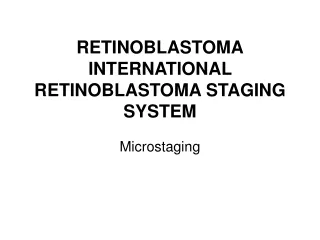
Major features of this classification • Separates conservatively treated patients from enucleated and metastatic ones • Proposes microstaging for putative risk factors of enucleated eyes • Extent of extraocular disease by imaging studies and pathology (e.g. CSF and BM)
Risk stratification according to stage Potential for cure Curability Stage 0 and 1 Stage 2 and 3 Stage 4
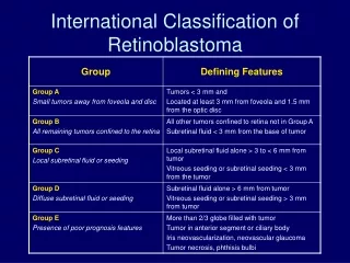
Information needed for treatment decisions • Post laminar optic nerve extension • Choroid invasion • Scleral invasion • Other ocular coats involvement (anterior segment) • Combination of these features
Optic Nerve microstaging • N0. No tumor in optic nerve • N1. Anterior lamina cribrosa • N2. Posterior lamina cribrosa • N3. Cut section and/or subarachnoid invasion • NX. Unknown
Choroid microstaging • C0. No choroidal invasion • C1. Superficial choroid invasion • C2. Deep choroid invasion
Scleral Microstaging • S0. No scleral involvement • S1. Microscopical extension into sclera • S2. Microscopical extension through sclera into the orbit
What will be this classification used for? • Having a standard for eye pathology and extent of disease evaluation • Comparing among different groups • Assessing incidence and disease extension by international registries • Defining the need for adjuvant therapy in special subgroups
Challenges • To define minimum standards for pathological processing of enucleated eyes • To prospectively validate the classification in a larger cohort • To disseminate it to groups and centers with high patient burden
Future steps • Interest eye pathologists • Set up definitions • Set up a registry • Provide a facility for centralized review for developing countries • e-teaching support • Analyze data to validate results