Download
1 / 21
210 likes | 317 Views
DNA Profiling and Biodiversity. Kath Crawford. Science and Plants for Schools, SSERC. DNA profiling and biodiversity. Thanks to:. Royal Botanic Garden, Edinburgh - Michelle Hollingsworth (Japanese knotweed) - Pete Hollingsworth (Epipactis youngiana)

E N D
DNA Profiling and Biodiversity Kath Crawford Science and Plants for Schools, SSERC
DNA profiling and biodiversity Thanks to: • Royal Botanic Garden, Edinburgh - Michelle Hollingsworth (Japanese knotweed) - Pete Hollingsworth (Epipactis youngiana) - Mark Hughes (Reforestation projects) • Scottish Initiative for Biotechnology (SIBE) Dr Jan Barfoot • University of Edinburgh Post-graduate science communication team
This morning’s activities Plant biodiversity through DNA profiling 09:00-09:30 Introduction Set up restriction digest 09:30-10:00 Restriction digest Demo of pouring gels 10:00-10:45 Loading wells, run gels Theory 10:45-11:05 Coffee 11:05-11:15 Stain gels 11:15 – 12:00 Roslin and SASA feedback 12:00-12:15 Observing gels 12:15-12:30 Closing remarks
This morning’s activities - aims • Offer hands-on experience of carrying out DNA restriction and electrophoresis suitable for use with post-16 students • Increase awareness and understanding of the use of DNA profiling in ecological applications • Simulate exercises in the use of DNA profiling in biodiversity applications • Explore how such practical work may support a Curriculum for Excellence
This morning’s activities Plant biodiversity through DNA Profiling three scenarios: • DNA profiling and Japanese knotweed (yellow microtubes) • DNA profiling and the characterisation of British orchids (blue microtubes) • DNA profiling and reforestation projects (green microtubes) Work in teams of six people, split into pairs, Each pair to carry out one scenario. At end, each pair to describe the scenario, results and any conclusions to the rest of the team of six.
Diagram: Dean Madden NCBE Using the microsyringes
Using the microsyringes • Never pull the plunger out of the microsyringe • Before loading, pull out the plunger a little (1 – 2 mm) • When dispensing liquid, hold the microsyringe as near to vertical as possible and at eye level • Remove the droplet of liquid from the end of the microsyringe tip by touching the inner wall of the microtube • Do not touch the point of the microsyringe tip with your fingers
Diagram: Dean Madden NCBE Dispensing the DNA sample • Put a clean tip on the microsyringe • Put 20 μL of G1, L, or K1 into tube containing dried restriction enzyme • Mix by drawing liquid up and down a few times even blue colour! • Cap tube tightly with a lid • Repeat steps above for other DNA samples • Put samples into a floating rack
Diagram: Dean Madden NCBE Incubation • Check that the tubes are firmly capped, then incubate them in a water bath at 37 ° C for 30 min Meanwhile, Anne and Gordon will demonstrate pouring gels
Diagram: Dean Madden NCBE Loading the gel • Your gel has been supplied covered with TBE buffer and with comb in place. • Very gently, ease the comb from the gel. • Put a clean tip on the microsyringe. Add 2 μL of loading dye to the tube containing DNA. Mix well by drawing the mixture up and down in the microsyringe tip.
Pipette the mixture of loading dye and DNA into one of the wells, holding the tip above the well but under the buffer solution. Take care not to puncture the bottom of the well! • Mark on the tank which DNA you have put into the well. • Repeat for your other DNA samples. Diagram: Dean Madden NCBE Loading the gel
Place gel tank in large electrophoresis tank and fit wires with crocodile clips Diagram: Dean Madden NCBE • Fit one electrode at each end of the tank. Running the gel
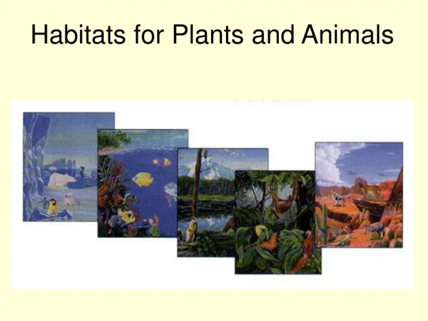
Epipactis youngiana Japanese knotweed - invasive, non-native species New species of orchid? Reforestation – which seed? © Christopher A Fields Japanese Knotweed growing through a pavement. Photo courtesy of Environment and Heritage Service, Cornwall. The scenarios
Japanese Knotweed Photo courtesy of Michelle Hollingsworth Japanese knotweed • Native to Japan, Taiwan, N China • Introduced to UK in 1800s as ornamental plant • Now widespread in British isles • - reproduces only asexually in Britain • - overruns British native plants • - causes damage • - difficult to eradicate • Categorised as invasive, non-native species and deliberate spread prohibited; classified as controlled waste • Important to understand genetics Fallopia japonica syn, Polyganum cuspidatum
Epipactis youngiana © Christopher A Fields Epipactis helleborine © Tim Rich Conservation of British orchids • Biodiversity action plans to honour ‘Convention on Biological Diversity’ • Epipactis youngiana – first described in 1980s • - found on mine spoil heaps in Northumberland and Glasgow • - thought to be new species • - given full conservation status • Limited resources for conservation purposes • Is E. youngiana a new species or a variant of E. helleborine?
Reforestation projects • Oak species are a major component of European forest resource • - provide habitat for other organisms • FAIROAK project to create map of oak genetic resources across Europe • - sampled ctDNA of oaks across Europe • - provided solid scientific information for use in development of conservation policies • Which seed to choose for reforestation project?
The practical - materials • Uses materials from the NCBE’s modular kits • - gel tanks, 6-tooth combs, microsyringes, calibrated microsyringe tips, carbon fibre electrode material, high-grade agarose, buffers, Azure A DNA stain • Uses restriction enzymes and DNA from the NCBE’s ‘Nature’s Dice’ kit • - BamH1 • - DNA
Diagram: Dean Madden NCBE The practical – the truth about the DNA samples • Three bacterial plasmids of different sizes • Plasmids mixed to give three DNA preparations (Mix 1, 2 and 3) • Each plasmid • - single site for BamH1 • - treatment with BamH1 cuts circular DNA to form a linear fragment that gives a single band after electrophoresis
Diagram: Dean Madden NCBE The practical – the truth about the DNA samples • DNA negatively charged • Gel porous • When voltage applied, DNA molecules move through gel towards positive electrode • DNA molecules separated by size
Pour ~ 10 mL Azure A stain on to the surface of the gel. Leave for exactly four minutes. Pour back into bottle. • Remove and dispose of electrodes. Pour off the buffer solution. Staining the gel
Diagram: Dean Madden NCBE Staining the gel • Rinse the surface of the gel very carefully with cold distilled or deionised water. Pour the water away. • Put the gel in a plastic bag to prevent it drying out, then leave to develop.