Download
1 / 28
590 likes | 1.49k Views
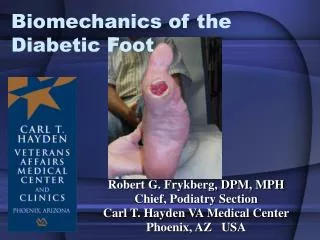
Biomechanics of the Diabetic Foot. Robert G. Frykberg, DPM, MPH Chief, Podiatry Section Carl T. Hayden VA Medical Center Phoenix, AZ USA. Diabetes Mellitus. Trauma. Neuropathy. Vascular Disease. MOTOR SENSORY AUTONOMIC MICROVASCULAR MACROVASCULAR

E N D
Biomechanics of the Diabetic Foot Robert G. Frykberg, DPM, MPH Chief, Podiatry Section Carl T. Hayden VA Medical Center Phoenix, AZ USA
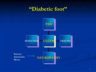
Diabetes Mellitus Trauma Neuropathy Vascular Disease MOTORSENSORYAUTONOMIC MICROVASCULARMACROVASCULAR Weakness Loss of Protective Anhidrosis Structural: Atherosclerosis Atrophy sensation Dry skin, Fissures Capillary BM Decreased Sympathetic thickening Deformity tone Functional: Ischemia (Altered blood flow A-V shunting Abnormal stress regulation) Increased blood flow Neuropathic edema High plantar pressure Callus formation Reduced nutrient capillary blood flow Osteoarthropathy Impaired Response to Infection Amputation Amputation DIABETIC FOOT ULCERATION RGF
Causal Pathways to Foot Ulcers High Plantar Foot Pressures Critical Triad in >63% of causal pathways From: Reiber et al: Diabetes Care 22:157-162, 1999
Altered Biomechanics in Diabetes • Biomechanical abnormalities / structural deformities are most frequently a consequence of Neuropathy • Altered gait patterns can result in unsteady gait with increased plantar foot pressures for longer durations (pressure-time integrals) • Combination of foot deformity and neuropathy increases the risk of ulcer • Limited Joint Mobility (ankle, STJ, great toe) will also lead to higher plantar pressures and ulcers Van Schie 2005 Cavanagh 1996
Contributing Factors to the Abnormal Mechanics of the Diabetic FootDiabetes MellitusNeuropathyStructural DeformityGait AbnormalitiesLJM Mononeuropathy Primary (idiopathic) Foot drop Collagen glycosylation Polyneuropathy Secondary Equinus reduced mobility Sensory Muscle atrophy Intrinsic muscle reduced shock absorption Motor Equinus atrophy increased pressures Autonomic Amputations Clawtoes Charcot Amputations Abnormal BiomechanicsHigh Plantar Pressures Neuropathic Ulceration Van Schie 2005 Zimny 2004 Frykberg 1995
Classification of Diabetic Neuropathy Generalized Symmetric Polyneuropathies • Acute Sensory • Chronic Sensorimotor • Autonomic Focal and multifocal neuropathies • Cranial • Truncal • Focal limb • Proximal motor (amyotrophy • Coexisting CIDP Boulton, Malik et al: Diabetes Care, 2004 Boulton, Vinik, et al: Diabetes Care, 2005
Intrinsic Muscle Atrophy Andersen et al: 2004
Intrinsic Muscle Atrophy Bus et al: Diabetes Care, 2002
Common Foot Deformities in Diabetes • Hammertoes (Clawtoes) • Bunions (hallux valgus) • Prominent metatarsal heads (pes cavus) • Charcot arthropathy • Partial foot amputations • Equinus (Achilles contracture) • Foot drop
STRUCTURAL DEFORMITY Primary (idiopathic)Pes cavus, pes planus, hallux valgus, hammertoes, forefoot deformities Deformities, pressure points, calluses precede neuropathy. Secondary"intrinsic minus foot"- clawtoes, pes cavus, depressed metatarsals. Loss of intrinsic muscle stability with long flexor over-dominance.Anterior crural atrophy (Ant. Tib.,EHL) - weakness, foot dropEquinus deformity- triceps surae dominance, post. tibial, long flexorsCharcot deformity - rocker bottom, Lisfranc subluxation, MTP subluxation Iatrogenic Post amputation: digital, ray, TMA, Lisfrancs, Choparts, Symes Frykberg 1995
AMPUTATIONS IN THE FOOTCONSEQUENCES • Structural alterations • Reduced contact areas • Increased plantar pressures • Altered function • Altered gait
Any deformity can lead to high plantar Pressures and subsequent ulceration in the Neuropathic Foot STRUCTURAL DEFORMITY Frykberg et al: J Foot Ankle Surg 2006
The Role of High Plantar Pressures in Diabetic Foot Ulceration • High plantar foot pressures are consistently detected in diabetic pts with neuropathy • Boulton 1987, Veves 1992, Stess 1997, Shaw 1998 • correlated with Limited Joint Mobility, plantar tissue thickness, and plantar fascia thickness • Zimny 2004, Abouaesha 2001, D’Ambrogi 2003 • risk factor for foot ulceration • Fernando 1991 Lavery 1998 Frykberg 1998 Lavery 2003 • Racial variations are evident • Veves 1995 Frykberg 1998
Predictive Value of Foot Pressure Assessment • 24 month study of 1666 DM patients • Mean age 69 yrs 50% male • Mean Duration DM 11.1 yrs • Mean Peak Plantar Pressure 86.6 N/cm2 • VPT 22.5 volts • 263 (15.8%) had or developed ulcer • Ulcer group had higher pressures Lavery LA, Armstrong, DG, et al, Diabetes Care 2003
Pressure is a factor Deformities IWGDF Foot Risk Categories Lavery LA, Armstrong, DG, et al, Diabetes Care 2003
Progressive Risk of Ulceration Neuropathy, PVD, And/ or Deformity No Neuropathy Neuropathy Hx Ulcer / Amp IWGDF Foot Risk Classification Peters 2001
GAIT DISTURBANCES • Function of neuropathy, deformity, & LJM • Abnormal loading patterns - earlier and longer • Altered cadence - instability and limp • Altered weight bearing sites – • Partial foot amputations - smaller area • Increased plantar pressures • Susceptible to ulceration
GAIT ABNORMALITIESContributing Factors • Proximal muscle atrophy - thigh weakness • Anterior crural atrophy - dorsiflexor weakness • Intrinsic muscle atrophy - clawtoes; reduced toe loading • Foot drop - Anterior tibial, Extensor hallucis longus paresis • Equinus - Posterior group dominance; triceps surae • Structural deformities - Charcot, post amputations
Limited Joint Mobility • A product of Nonenzymatic glycosylation of collagen • Also associated with retinopathy • Decreased ankle and hallux motion • Restricted subtalar range of motion • reduced shock absorption; • Increased vertical and shear forces • Increased peak plantar pressures • Alone does not cause ulceration • With neuropathy, contributes to plantar ulceration Delbridge 1988 Fernando 1991 Zimny 2004
The Role of Limited Joint Mobility in Diabetic Patients with an At-Risk FootZimny, Schatz, Pfohl: Diabetes Care 27:942-946, 2004
There is a strong inverse correlation between joint mobility and PTI in diabetic patients At-Risk Neuropathic patients have less joint mobility and higher PTI’s than control DM (non-neuropathic) patients Zimny: Diabetes Care, 2004
Equinus Deformity • Achilles tendon contracture • Increases plantar forefoot pressure • May increase risk for ulceration • Present in ~ 40% of high-risk patients • At 3x greater risk for presenting with high plantar pressures Barry DC et al, JAPMA, 1993 Grant WP et al, JFAS, 1997 Lavery, et al, Arch Intern Med, 1998 Lavery, Armstrong, Boulton, JAPMA, 2002
Equinus Deformity • Diabetic population study San Antonio, TX n=1666 • 50% male Age ~ 69 yrs • Duration DM 11.1 yrs • VPT ~ 22.5 • Equinus Prevalence 10.3% • Peak plantar pressure 86.6 N/cm2 Lavery, Armstrong, Boulton, JAPMA, 2002
Biomechanics of The Diabetic Foot • Biomechanical alterations are a composite function of neuropathy, structural deformity, LJM, and associated gait disturbances • Neuropathy is a primary determinant • Early recognition, intervention, and prevention of deformity with high plantar pressures are crucial to the avoidance of ulceration
You can observe a lot just by watching Yogi Berra American Baseball Player and Philosopher
THANK YOU! Robert G. Frykberg, DPM, MPH Robert.frykberg@med.va.gov