Download
1 / 49
1.18k likes | 4.49k Views
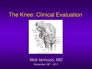
Clinical Anatomy of the knee. Mr CM Gupte Mr Alvin Chen. Overview. Knee joint function Surface anatomy Bones Ligaments Tendons Examination Disease processes. The Knee Joint. Poorly constructed in terms of stability - femur round, tibia flat. Comprised of four bones. Femur Tibia

E N D
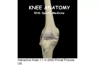
Clinical Anatomy of the knee Mr CM Gupte Mr Alvin Chen
Overview • Knee joint function • Surface anatomy • Bones • Ligaments • Tendons • Examination • Disease processes
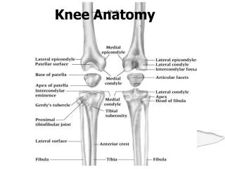
The Knee Joint • Poorly constructed in terms of stability - femur round, tibia flat. • Comprised of four bones. • Femur • Tibia • Fibula • Patella
The knee joint • Load bearing / Force transmission • Locomotion • Proprioception
Bones • Patella: femur = patellofemoral joint • Femur: tibia = medial and lateral tibiofemoral joints
Patella • Medial facet • Lateral facet • Articular cartilage
Femur • Medial femoral condyle • Lateral femoral condyle (protrudes more) • Trochlea • Articular cartilage
Femoral ligament insertions • PCL inner aspect medial femoral condyle • ACL inner aspect lateral femoral condyle
Tibia • Medial and lateral plateau • Insertions of menisci and cruciate ligaments • MCL/semimembranosus
Tibial plateau • Insertions of menisci/ • Cruciate ligaments
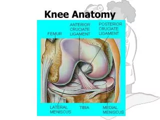
Ligaments • Extracapsular: MCL/ LCL • Intracapsular: ACL/PCL Functions: • Stabilise • Proprioception
ACL • Two bundles • Prevents anterior drawer and pivot
MRI Normal ACL in RED Torn ACL
Tests for ACL Lachman’s Anterior Draw
Arthroscopy Intact ACL Torn ACL
PCL • Stronger than ACL • Prevents posterior drawer
PCL injury • Direct Blow onto Tibia on a flexed knee
MRI PCL Normal PCL PCL torn
MCL • Medial • Prevents valgus stress • Deep and superficial parts • Heals well
LCL • Lateral • Prevents varus stress • Cord like –weakest of the ligaments but rarely torn in isolation
Extensor mechanism • Quadriceps • Patella • Patellar tendon
Quadriceps • VMO • Rectus femoris • Vastus intermedius • Vastus lateralis • Extend knee
VMO • Medial most quad • Helps prevent lateral dislocation of the patella
Hamstrings • Biceps laterally • Semitendinosus/semimembranosus medially • Flex knee
Pes • Sartorius • Gracilis • Semitendinosus • G and T used for ACL reconstruction
Movements of the Knee The principal muscles acting on the knee: • Extensors - quadriceps femoris • Flexors - hamstrings assisted by gracilis, gastrocnemius and sartorius. • Medial rotators- popliteus.
Movements of the Knee • The principal knee movements are flexion and extension, but rotation of the knee is possible when the joint is in flexed position .
Examination • Look • Feel • Move • Think about structures and what you’re dong to them
Diseases • OA • Meniscal tears • Ligament injuries • Patellofemoral tracking • Tendon injuries • Inflammatory arthritis • Infection/tumours
Treatment of Meniscal Tears Suture/ Repair Debridement
Other Pathologies Patella Maltracking Bone Tumour