Download
1 / 28
280 likes | 522 Views
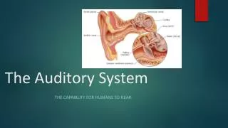
Central Auditory System. I. Tonotopic organization (i.e . frequency map in the central auditory system) - Sound is represented tonotopically in the cochlea, which results in tonotopy in the auditory nerve fibers. .

E N D
I. Tonotopicorganization • (i.e. frequency map in the central auditory system) • - Sound is represented tonotopically in the cochlea, which results in tonotopy in the auditory nerve fibers.
Auditory nerve fibers that innervate the basal end of the basilar membrane are tuned to high frequencies and fibers that innervate the apex are tuned to low frequencies; the frequency to which a particular auditory neuron is most sensitive is called its best frequency (BF).
- Tonotopy is preserved throughout central auditory system, e.g. as evident in a Golgi stain of the cochlear nuclei • - The mapping of frequency into place in the auditory system corresponds to the perceptual sense of lowness or low pitch (low frequency) and highness or high pitch (high frequency).
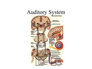
II. Neuroanatomy of the auditory system • - Sequence • (1) Auditory nerve fibers from cochlea terminate in cochlear nuclei (DCN or VCN) • (2) From the CN, most neurons project to superior olivary nuclei for sound localization.
The two principal nuclei the medial (MSO) and lateral (LSO) receive synaptic binaural inputs from bushy cells of the VCN. • The SO is the site of initial computations for sound localization.
(3) The lateral lemniscus is a collateral path to the inferior colliculus • (4) All ascending neurons (from SO and LL) terminate in the inferior colliculus • (5) IC projects to the medial geniculate (auditory thalamus) • - Most commisures occur at the trapezoid body, and above the SO most neurons are bi-aural.
III. Sound localization by azimuth • - Locating a sound source carries the additional problems of • (1) distinguishing multiple sound sources over background noise and • (2) suppressing the effect of echoes.
- Due to path length difference, a sound source will have different effects on the right and left ears • (1) time difference: interaural time difference (ITD) and interaural phase difference (IPD) • (2) loudness difference: interaural level difference (ILD)
- The ITD has at least two aspects: • 1) envelope ITD, meaning delay of the envelope of the stimulus • (e.g. the onset and offset times and the times of any amplitude fluctuations). • 2) IPD, the delay of the actual stimulus waveform, most easily expressed as the delay of the zero-crossings or waveform peaks. • IPD is better perceived because it is ongoing throughout the sound and its phase-locking of ANFs.
- Relative importance of ITD and ILD cues depends on stimulus frequency • - The superior olivary nuclei compute sound localization. • Neurons in the MSO receive terminals from specialized cells called bushey cells in the cochlear nuclei of both sides.
Neurons in LSO receive a similar symmetric innervation, except the projection from the contralateral ear passes thru an inhibitory interneuron in the medial nerve of the trapezoid body (MNTB), which is another component of the superior olive.
Busheycells receive large specialized synapses from auditory nerve (AN) fibers on their somata. • These synapses may contain hundreds of individual synaptic contacts and constitute a very powerful input to the bushy cell. • Bushy cells are further specialized in that their postsynaptic membranes contain a relatively high conductance K+ channel which is activated at potentials just depolarized from rest.
Because of this channel, bushy cell time constants are fast so that these cells can follow individual synaptic events without temporal summation. • The combo of the large synaptic input from AN fibers and the short time constant allows bushy cells to preserve waveform timing (phase-locking) information from AN fibers.
The timing info is needed to support ITD calculations in MSO neurons. • - MSO neurons measure the ITD • - MSOs are activated by coincident stimulation by both ears • - The ITD to which a MSO is most sensitive depends on the internal neural delay (function of path length of bushy cells on the two sides).
By varying the internal neural delay in the bushy cell circuits, a range of ITD sensitivities can be produced in MSO neurons. • - LSO neurons measure the ILD • - LSO neurons are stimulated when ipsilateral intensity is greater than contralateralintensity.
IV. Sound localization by elevation • - It is more difficult to determine elevation of cue; “cone of confusion” is apparent in single tone stimulus • - Sound reflections off the pinna differ depending on elevation of sound source; the resulting interference causes nulls at high frequency, and the location of the null is a function of elevation.
V. Auditory (cortex) neurons may encode stimuli through coordinated firing in response to a stimulus • VI. Descending systems • 1 - Nerves to the middle ear muscles (stapedius (CN VII) and tensor tympani (CN V)) dampen effect of loud sounds and help filter out low-frequency self-generated sounds (e.g. due to chewing) and thus maintaining sensitivity of the system for external sounds.
2 - Projections from the olivocochlear bundle (OCB) is a pair of projections from cell bodies in the SO to the cochlea. • MSO project thru the crosses component (COCB) it innervate outer hair cells; neurons near LSO project thru the uncrossed component (UCOCB) to innervate AN dendrites near inner hair cells. • Electrical stimulus of the COCB near the floor of the 4th ventricle produces a substantial suppression of the response of IHCs.
A similar suppression is observed in the AN fibers contacting the IHCs. • This suppression occurs without a direct neural connections (instead, via OHCs) by modifying the mechanical response of the organ of Corti which drive the IHCs.
The role of the OCB appears to be to improve the ability of the auditory system to discriminate the vowels /i/ and /u/ (and general hearing?) in the presence of background noise without affecting their ability in quiet.
Neural Substrates of Language: Evidence from Aphasia. • Telegraphic – prepositions and conjunctions are omitted. • The hallmark of Broca’s aphasia is nonfluent, agrammatic speech output. • Spoken output is limited to content words that do not form a grammatical sentence.
Comprehension is relatively spared, but there is difficulty with syntactically complex sentences. • Naming is generally impaired. • Often nouns are names more correctly than verbs. • Damage to Broca’s area causes selective impairment in planning and executing the complex movements to articulate words (aphemia).
Wernicke’s • English-like but empty in meaning, may use neologisms • Characterized by prominent impairment in understanding spoken words and sentences. • In addition, while words and word-like utterances are produced effortlessly and with the basic structure and melody of sentences, the link between the words and their meanings seems to be disrupted.
Disproportionally impaired word and sentence repetition. • Speech is fluent and grammatical, although phonemic paraphasias are often present. • “Conduit d’aaproche” is the frequently observed phenomenom of progressive self-correction of speech errors.
Left ACA or watershed between ACA and MCA • Comprehension and content of speech are relatively intact, while fluency and articulation are impaired in spontaneous speech but not in sentence repetition.
Watershed between MCA and PCA • Comprehension of language and content of speech are markedly impaired, but sentence repetition is relatively accurate.
Comments on correlates with lesion sites and vascular supply • - Best correlations are with specific language tasks and lesion sites (not aphasia syndrome and lesion site) • - Some language tasks, like picture identification, are performed by a network of relatively discrete regions of the cortex; lesions at any part of the network may produce a defect in that language task.