Download
1 / 44
650 likes | 1.15k Views
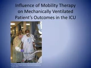
Monitoring the Mechanically Ventilated Patient. Mazen Kherallah, MD, FCCP Critical Care Medicine and Infectious Disease. Monitoring in the Past. Visual monitoring of respiration and overall clinical appearance Finger on pulse Blood pressure (sometimes).

E N D
Monitoring the Mechanically Ventilated Patient Mazen Kherallah, MD, FCCP Critical Care Medicine and Infectious Disease
Monitoring in the Past • Visual monitoring of respiration and overall clinical appearance • Finger on pulse • Blood pressure (sometimes)
Harvey Cushing Not just a famous neurosurgeon … but the father of anesthesia monitoring • Invented and popularized the anesthetic chart • Recorded both BP and HR • Emphasized the relationship between vital signs and neurosurgical events( increased intracranial pressure leads to hypertension and bradycardia )
Monitoring in the Present • Standardized basic monitoring requirements (guidelines) from the ASA (American Society of Anesthesiologists), CAS (Canadian Anesthesiologists’ Society) and other national societies • Many integrated monitors available • Many special purpose monitors available • Many problems with existing monitors (e.g., cost, complexity, reliability, artifacts)
Low Tech Patient Monitoring • Manual blood pressure cuff • Finger on the pulse and forehead • Monaural stethoscope (heart and breath sounds) • Eye on the rebreathing bag (spontaneously breathing patient) • Watch respiratory pattern • Watch for undesired movements • Look at the patient’s face • color OK? • diaphoresis present? • pupils
High Tech Patient Monitoring Examples of Multiparameter Patient Monitors
High Tech Patient Monitoring Transesophageal Echocardiography Depth of Sedation Monitor
Airway / Respiratory Axis • Correct ETT placement • ETT cuff pressure • Suctioning • Oxygenation • Ventilation • Airway pressure • Airway gas monitoring • Clinical: wheezing, crackles, equal air entry, color, respiratory pattern (rate, rhythm, depth, etc.)
Correct ET Tube Placement:Capnography Purpul Yellow
Correct ET Tube Placement • Secure ET tube in place, note the number • Sedate patient with appropriate MAAS • Avoid accidental, or self extubation
Tracheal Tube Cuff Care • These include bedside sphygmomanometers, special aneroid cuff manometers, and electronic cuff pressure devices. • Ideally, most tubes seal at pressures between 14 and 20 mm Hg (19 to 27 cm H2O). • Tracheal capillary pressure lies between 20 and 30 mm Hg • Impairment in tracheal blood flow seen at 22 mm Hg and total obstruction seen at 37 mm Hg
Minimum Leak Volume Technique • Air inflation of the tube cuff until the airflow heard escaping around the cuff during positive pressure breath ceases. • Place a stethoscope over larynx. Indirectly assesses inflation of cuff. • Slowly withdraw air (in 0.1-mL increments) until a small leak is heard on inspiration. • Remove syringe tip, check inflation of pilot balloon
Beers Law for Dummy’s What is the amount of light absorbed by the “peak” of the cardiac cycle
Conditions Affecting Accuracy • Patient conditions • Carboxyhemoglobin • Erroneously high reading may present • Anemia • Values as low as 5 g/dl may result in 100% SpO2 • Hypovolemia/Hypotension: • May not have adequate perfusion to be detected by oximetry • Hypothermia: • peripheral vasoconstriction may prevent oximetry detection
Patient Environments • Ambient Light • Any external light exposure to capillary bed where sampling is occurring may result in an erroneous reading • Excessive Motion • Always compare the palpable pulse rate with the pulse rate indicated on the pulse oximetry • Fingernail polish and pressed on nails • Most commonly use nails and fingernail polish will not affect pulse oximetry accuracy • Some shades of blue, black and green may affect accuracy (remove with acetone pad) • Skin pigmentation • Apply sensor to the fingertips of darkly pigmented patients
Endotracheal suctioning: Indications • Coarse breath sounds • Noisy breathing • Visible secretions in the airway • Decreased SpO2 in the pulse oximeter & deterioration of arterial blood gas values • Clinically increased work of breathing • Suspected aspiration of gastric or upper airway secretions • Changes in monitored flow/pressure graphics • Increased PIP; decreased Vt during ventilation
Endotracheal or Tracheostomy Tube Suctioning Open Suctioning Disconnection from the ventilator Not recommended when PEEP >10 Closed Suctioning: Facilitate continuous mechanical ventilation and oxygenation during the suctioning. Indicated when PEEP level above 10cmH2O
High Minute Volume alarm • Fever • Sepsis • Hyperthyroidism • Agitation • Overfeeding
Static and Dynamic Pressures Pressure PIP Flow-Resistive Pressure difference (Pres) Pplat Alveolar Distending (recoil) Pressure difference (Pdis) PEEP time
Peak Inspiratory Pressure Plateau Pressure
High PIP Normal PPlat
High PIP High PPlat
Inspiratory Plateau Pressure • Pneumothorax • Hemothorax • Pleural effusion • Pneumonia • Congestive heart failure • ARDS
Persistant Flow at end expiration • Auto PEEP: Dynamic Hyperinflation • Management: • Decrease Tidal Volume • Decrease Insp/Exp ratio • Increase inspiratory flow • Decrease rate • Decrease Inspiratory time
Low exhaled volume • System leak: Gurgling sound in the neck area • Dislodged ETT • Deflated cuff • Punctured cuff • Bronchopleural fistula: Gurgling sound in the chest area
If you are admitted to our ICU and intubated we will: • Secure your ETT and avoid accidental or self extubation • Will monitor your cuff pressure to avoid tracheal wall pressure injury • Suction you with closed system • Set ventilator alarms to detect variations early • Elevate the head of bed to > 30º • Use deep venous thrombosis prophylaxis when indicated • Use Stress ulcer prophylaxis
Thank you Mazen Kherallah, MD, FCCP Critical Care Medicine and Infectious Disease