Download
1 / 74
900 likes | 1.53k Views
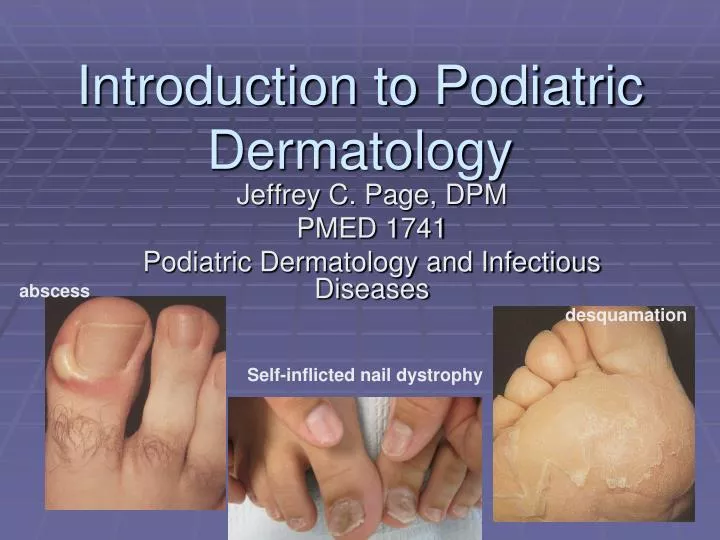
Introduction to Podiatric Dermatology. abscess. desquamation. Jeffrey C. Page, DPM PMED 1741 Podiatric Dermatology and Infectious Diseases. Self-inflicted nail dystrophy. Introduction to Podiatric Dermatology. General Principles Skin anatomy Lesion types Patterns & Configurations

E N D
Introduction to Podiatric Dermatology abscess desquamation Jeffrey C. Page, DPM PMED 1741 Podiatric Dermatology and Infectious Diseases Self-inflicted nail dystrophy
Introduction to Podiatric Dermatology • General Principles • Skin anatomy • Lesion types • Patterns & Configurations • Disorders of Sweat • Mechanical Lesions Exfoliation from reaction to gluten Depigmentation post steroid injection
Skin Anatomy Layers Appendages Purpose
Epidermal Layers • Stratum corneum • Stratum lucidum (palms and soles) • Stratum granulosum • Stratum spinosum (prickle cell layer) • Stratum basale (germinativum) • Merkel’s cells (storage granules) • Langerhans’ cells (immune system) • Melanocytes (produce melanin) • Keratinocytes (produce keratin)
Dermal Layers • Dermal-epidermal junction = basement membrane • Papillary dermis (superficial) • Reticular dermis (deep) • Vessels • Nerves • Appendages (adnexa) • Hair • Glands (sweat & sebaceous) • Nails
Subcutaneous Layer • Production and storage of fat cells • Supports blood vessels and nerves • Storage of nutrition • Thermoinsulation
Description of Lesions • Primary Lesions • Secondary Lesions • Elementary Lesions melanoma atrophy from injections
Descriptions Must Include: • Lesion type/ texture • Size/area • Moisture level • Color • Location
Macules Patches Papules Plaques Nodules Wheals Vesicles Bullae Pustules Cysts Tumors Primary Lesions
Macules & Patches Lentigo Hyperpigmentation from venous stasis Hypopigmentation from skin trauma
Papules verrucae nevus Kaposi’s sarcoma
Plaques psoriasis tinea corporis/pedis
Nodules Neurofibromatosis Nodular melanoma Hemangioma
Vesicles & Bullae Vesicular tinea Chicken Pox herpes
Wheals (Hives) truncal placques dermatographism angioedema
Cysts* & Tumors Inclusion cyst Pedunculated nevus *Cysts are sometimes called special lesions
Scales Crusts Excoriations Fissures Ulcers Erosions Scars Induration Atrophy Lichenification? Secondary Lesions Plantar ulcer
Scales Icthyosis Severe edema due to heart failure Atopic dermatitis
Crusts Impetigo Scalp kerion
Erosions & Ulcers osteomyelitis Circinate balanitis Syphilitic chancre
Fissures & Atrophy Fissure from frequent dishwashing Atrophy and telangiectasia due to oral steroids
Scars keloid Hypertrophic scar following buionectomy Scars in construction worker Hypopigmented scar from acne
Elementary (Special) Lesions • Excoriations • Comedones • Burrows • Purpura • Petechiae • Ecchymoses • Macular or Papular • Telangiectasia • Lichenification? • Cyst? Sebacious cyst
Elementary (Special) Lesions Excoriations in factitial dermatitis Comedones
Purpura Vasculitides
Elementary (Special) Lesions Burrow in Scabies Telangiectasia from sun exposure
Elementary (Special) Lesions Spider Angioma Lichenification
Round Annular Discoid Targetoid Nummular Arciform-polycyclic Geographic Herpetiform-grouped Serpiginous-gyrate Linear Reticular Verrucous Guttate Patterns and Configurations
Round Lesions Dermatofibroma Tinea pedis/corporis Discoid Annular
Round Lesions Erythema multiforme Nummular eczema Targetoid Nummular
Arciform (polycyclic) Lichen planus
Geographic Lesions Pyoderma gangrenosum Pitted keratolysis Necrobiosis lipoidica diabeticorum melanoma
Herpetiform (Grouped) Lesions Herpes zoster Herpes zoster
Serpiginous (Gyrate) Poison ivy Leishmaniasis
Linear Lesions scratches wheals