Download
1 / 1
10 likes | 140 Views
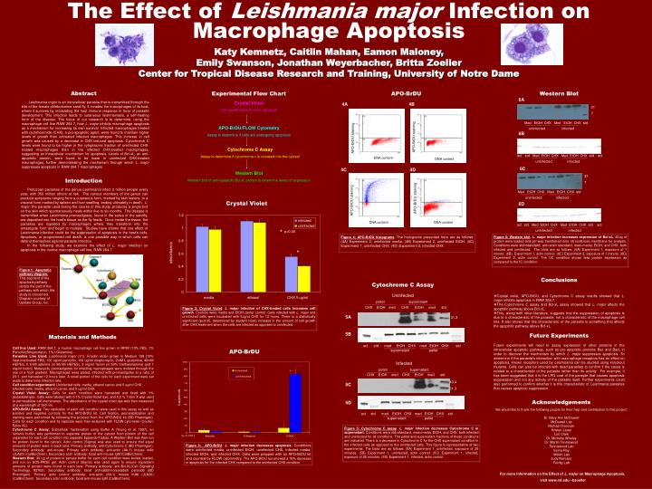
The Effect of Leishmania major Infection on Macrophage Apoptosis Katy Kemnetz, Caitlin Mahan, Eamon Maloney, Emily Swanson, Jonathan Weyerbacher, Britta Zoeller Center for Tropical Disease Research and Training, University of Notre Dame. Abstract. Experimental Flow Chart. APO-BrDU.

E N D
The Effect of Leishmania major Infection on Macrophage Apoptosis Katy Kemnetz, Caitlin Mahan, Eamon Maloney, Emily Swanson, Jonathan Weyerbacher, Britta Zoeller Center for Tropical Disease Research and Training, University of Notre Dame Abstract Experimental Flow Chart APO-BrDU Western Blot 6A Leishmania major is an intracellular parasite that is transmitted through the bite of the female phlebotomine sand fly. It invades the macrophages of its host, where it survives by modulating the host immune response in favor of parasite development. This infection leads to cutaneous leishmaniasis, a self-healing form of the disease. The focus of our research is to determine, using the macrophage cell line RAW 264.7, how L. major inhibits macrophage apoptosis as a mechanism for increasing its own survival. Infected macrophages treated with cycloheximide (CHX), a pro-apoptotic agent, were found to maintain higher levels of growth than untreated infected macrophages. This increase in cell growth was caused by a decrease in CHX-induced apoptosis. Cytochrome C levels were found to be higher in the cytoplasmic fraction of uninfected CHX-treated macrophages than in the infected CHX-treated macrophages, suggesting an intracellular mechanism for apoptosis. Levels of Bcl-xL, an anti-apoptotic protein, were found to be lower in uninfected CHX-treated macrophages, further demonstrating the mechanism through which L. major suppresses apoptosis in RAW 264.7 macrophages. Crystal Violet Cell growth assay to infer cell death 4A 4B 31 7 Med EtOH CHX Med EtOH CHX std APO-BrDU/FLOW Cytometry Assay to determine if cells are undergoing apoptosis uninfected infected 6B APO-BrDU staining APO-BrDU staining Cytochrome C Assay Assay to determine if cytochrome c is released into the cytosol act std Med EtOH CHX Med EtOH CHX std act DNA content DNA content uninfected infected 6C 4C 4D Western Blot Western blot of anti-apoptotic Bcl-xL protein to determine levels of expression 31 7 Introduction Protozoan parasites of the genus Leishmania infect 2 million people every year, with 350 million others at risk. The various members of the genus can produce symptoms ranging from a cutaneous form, marked by skin lesions, to a visceral form marked by spleen and liver swelling, ending ultimately in death. L. major, the parasite used during the course of this study, produces a single boil on the skin which spontaneously heals within five to six months. The disease is transmitted when Leishmania promastigotes, found in the saliva of the sandfly, are deposited into the host’s tissue as the fly feeds. Once inside the tissue, the parasites are ingested by macrophages where they transform into the amastigote form and begin to multiply. Studies have shown that one effect of Leishmania infection could be the suppression of apoptosis in the host’s cells. Apoptosis, or programmed cell death, is one possible way in which cells can defend themselves against parasite infection. In the following study, we examine the effect of L. major infection on apoptosis in the murine macrophage cell line, RAW 264.7. Med EtOH CHX Med EtOH CHX std APO-BrDU staining uninfected infected Crystal Violet APO-BrDU staining 6D DNA content DNA content act std Med EtOH CHX Med EtOH CHX std act * uninfected infected p<0.05 Figure 4: APO-BrDU histograms. The histograms presented here are as follows: (4A) Experiment 2, uninfected media. (4B) Experiment 2, uninfected EtOH. (4C) Experiment 1, uninfected CHX. (4D) Experiment 2, infected CHX. Figure 6: Western blot. L. major infection increases expression of Bcl-xL. 30ug of protein were loaded and gel was transferred onto nitrocellulose membrane for analysis. Conditions were std=standard, act=actin standard,med=media, EtOH, and CHX, both infected and uninfected. The blots are as follows: (6A) Experiment 1, exposure of 1 minute. (6B) Experiment 1, actin control. (6C) Experiment 2, exposure of 1 minute. (6D) Experiment 2, actin control. The UC condition shows less protein expression as compared to the IC condition. absorbance * Figure 1: Apoptotic pathway diagram. This segment of the apoptosis pathway details the part of the pathway with which this study is concerned. Diagram courtesy of Upstate Group, Inc. Conclusions Cytochrome C Assay • Crystal violet, APO-BrDU, and Cytochrome C assay results showed that L. major inhibits apoptosis in RAW 264.7. • The Cytochrome C assay and Bcl-xL assay showed that L. major affects the apoptotic pathway above Bcl-xL. • This, along with other literature, suggests that the suppression of apoptosis is due to a characteristic of the parasite, not a characteristic of the macrophage cell line. It also shows that this characteristic of the parasite is something that affects the apoptotic pathway above Bcl-xL. • Future Experiments • Future experiments will need to assay expression of other proteins in the intracellular apoptotic pathway, such as pro-apoptotic proteins Bax and Bad, in order to discover the mechanism by which L. major suppresses apoptosis. To determine if the parasite’s interaction with macrophage receptors has an effect on apoptosis, known receptors used by Leishmania can be studied using knockout mutants. Cells can also be infected with dead parasites to confirm if the cause is related to a characteristic of the parasite rather than its activity. For example, it has been suggested that it is the LPG coat of the parasite that causes apoptosis suppression and not any activity of the parasite itself. Further experiments could also performed to confirm whether it is this characteristic of Leishmania parasites that causes apoptotic suppression. Uninfected pellet supernatant CHX EtOH med CHX EtOH med std Figure 2:Crystal Violet. L. major infection of CHX-treated cells increases cell growth. Controls were media and EtOH carrier control. Cells infected with L. major and uninfected cells were incubated with 5ug/ul CHX for 12 hours. There is a statistically significant (p<0.05, determined by student t-test) increase in the amount of cell growth after CHX treatment when the cells are infected as opposed to uninfected. 5A 31.3 Materials and Methods 5B act std med EtOH CHX med EtOH CHX std Cell line Used: RAW 264.7, a murine macrophage cell line grown in RPMI (10% FBS, 1% Penicillin/Streptomycin, 1% l-Glutamine). Parasites Line Used: Leishmania major (V1), Friedlin strain grown in Medium 199 [10% heat-inactivated FBS, 100 ug/ml penicillin, 100 ug/ml streptomycin, 2mM L-glutamine, 40mM HEPES, 0.1mM adenine (in 50mM HEPES), 5 mg/ml hemin (in 50% triethanolamine) and 1 mg/ml biotin]. Metacyclic promastigotes for infecting macrophages were isolated through the use of a ficoll gradient. Macrophages were plated, infected with promastigotes at a ratio of 20:1, and harvested 12 hours later. A small portion of the cells for each experiment were set aside to determine infection rate. Cell condition/experiment: Uninfected cells: media, ethanol carrier and 5 ug/ml CHX Infected cells: media, ethanol carrier, and 5 ug/ml CHX. Crystal Violet Assay: Cells for each condition were harvested and fixed with 1% glutaraldehyde. Cells were labeled with 0.1% Crystal Violet dye, and 0.2 % Triton X was used to permeabilize cell membranes. The absorbance of the crystal violet dye was then measured at a wavelength of 590 nm. APO-BrDU Assay: Two replicates of each cell condition were used in this assay as well as positive and negative controls for the APO-BrDU kit. Cell fixation, permeabilization and staining were performed by following the protocol from the APO-BrDU kit (BD Pharmigen). Cells for each condition and its replicate were then analyzed with FLOW cytometer (Coulter, Epics XL). Cytochrome C Assay: Subcellular fractionation using Buffer A (Young et al, 1997), an isotonic buffer, was performed to separate protein of the cytosol from protein of the cell organelles for each cell condition into separate Eppendorf tubes. A Western Blot was then run for protein found in the cytosol. Actin control (Sigma) was also used to ensure that equal amounts of protein were in each lane. Primary antibody: anti-Cytochrome C (BD Pharmigen). Secondary antibody: anti-mouse. Primary actin antibody: anti-actin (Ab-1) mouse mAb (JLA20) (CalBioChem). Secondary actin antibody: Goat anti-mouse IgM (CalBioChem). Western Blot: 30 ug of protein in sample buffer for each cell condition were boiled, loaded, and run on SDS-PAGE gel. Actin control (Sigma) was used again to ensure equivalent amounts of protein were found in each lane. Primary antibody: anti-Bcl-XL(Cell Signaling Technology, #2762). Secondary antibody: Goat anti-rabbit-horseradish peroxide (BD Pharmigen). Primary actin control antibody: anti-actin (Ab-1) mouse mAb (JLA20) (CalBioChem). Secondary actin antibody: Goat anti-mouse IgM (CalBioChem). APO-BrDU supernatant pellet Infected pellet supernatant CHX EtOH med CHX EtOH med std 40.4 31.3 5C Acknowledgements 5D We would like to thank the following people for their help and contribution to this project: Dr. Mary Ann McDowell McDowell Lab Michael Donovan Kristen Leary Lori Clark Dr. Michelle Whaley Dr. Martin Tenniswood Tenniswood Lab Soma Roy Welsh Lab Judy Narvaez Ferdig Lab act std med EtOH CHX med EtOH CHX std supernatant pellet Figure 5: Cytochrome C assay.L. major infection decreases Cytochrome C in supernatant. Conditions were std=standard, med=media, EtOH, and CHX, both infected and uninfected for all conditions. The pellet and supernatant fractions of these conditions are indicated. There is a decrease in Cytochrome C for the CHX supernatant condition in the infected cells as opposed to the uninfected cells. This figure is representative of two experiments. The blots are as follows: (5A) Experiment 1, uninfected, exposure of 25 minutes. (5B) Experiment 1, uninfected, actin control. (5C) Experiment 1, infected, exposure of 25 minutes. (5D) Experiment 1, infected, actin control. Figure 3: APO-BrDU. L. major infection decreases apoptosis. Conditions were uninfected media, uninfected EtOH, uninfected CHX, infected media, infected EtOH, and infected CHX. Cells were prepped with an APO-BrDU kit and counted by FLOW cytommetry.The APO-BrDU run showed a 76% decrease in apoptosis for the infected CHX compared to the uninfected CHX condition. For more information on the Effect of L. major on Macrophage Apoptosis, visit www.nd.edu/~bzoeller