Download
1 / 63
630 likes | 796 Views
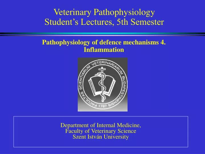
Veterinary Pathophysiology Student’s Lectures, 5th Semester. Pathophysiology of defence mechanisms 4. Inflammation. Department of Internal Medicine, Faculty of Veterinary Science Szent István University. DEFENCE MECHANISMS DEVELOPED DURING THE PHYLOGENESIS (Handouts p 28).

E N D
Veterinary PathophysiologyStudent’s Lectures, 5th Semester Pathophysiology of defence mechanisms 4. Inflammation Department of Internal Medicine, Faculty of Veterinary Science Szent István University
DEFENCE MECHANISMS DEVELOPED DURING THE PHYLOGENESIS (Handouts p 28)
HISTORICAL BACKGROUND Aulus Cornelius Celsus (1.század): "De Re Medicina": rubor (redness), dolor (pain), calor (heat), tumor (swelling) Galenus(AD 130-200), Virchow(1821-1902): functio laesa (loss of function) Hunter, Cohenheim: importance of vascular factors Mechnicov: phagocytosis Dale, Laidlow, Lewis: histamine - notion of "mediators" Schwartzmann: endotoxin caused inflammation Arthus: Immune Complex Mediated Hypersensitivity (Type III.) Menkin: discovered ancestors of the "peptid mediators" Menkin`s "exudine"=bradykinin Ratnoff: significance of Hageman (XII.)-factor De Duve: lysosomes
Ex Libris of The International Club of Inflammation CalorRubor Tumor Dolor Functio laesa
Three major parts of the inflammatory response: • Hemodynamic changes • Permeability changes • Cells involved
Hemodynamic changes • Transient vasoconstriction: lasts for a few seconds - neurogenic mechanism • Vasodilation: begins in a few minutes and lasts hours or longer, release of histamine form mast cells, generation of nitric oxide (NO) and prostaglandins (PGD2, PGE2) – causing acute local active hyperemia • Increased volume of blood flow in the area (a result of vasodilation) • Increased vascular permeability (histamine, leukotrienes C4, D4, E4, bradykinin, and platelet activating factor (PAF) cause contraction of endothelial cells resulting gaps in the small vessels. Complement factors (C5a and C3a) indirectly participate by inducing release of histamine by mast cells. • Rate/speed of flow decreases (caused by vasodilation in the area) • Margination of leukocytes along vessel walls: a decreased rate of flow results in decreased shear force on white blood cells (WBC) as they contact the vessel wall. • Exudation: WBCs migrate out into the tissue as does the serum.
Permeability changes Vascular leakage after local injury can occur: 1. directly; as an effect of any kind of structural injury to the microvasculature affecting all types of vessels. 2. indirectly; as an effect of biochemical susbtances that appear in and around the site and affect primarily the venules. Permeability can be: 1. passive process - passage of fluid between the endothelial cells; 2. active process - endothelial cells create endocytotic vesicles, and discharge the contents on the other side by active transport within the cell istelf. Development of edema can occur by two major mechanisms: 1. purely hydrostatic; 2. permeability effected by biochemical mediators.
Permeability changes Exudation
Permeability effected by biochemical mediators: Biochemical mediators are stored and released from cells (ie. histamine and serotonin from mast cells, basophil granulocytes and platelets), or can be synthesized rapidly. Plasma kinins (bradykinin - activated or cleaved enzymatically by plasma proteinases including Hageman factor /XIIa/) Complement factors (C5a, C3a - induce mediator release from leukocytes Leukotrienes (LTC4, LTD4, LTE4, LTB4 - produced through the arachidonic acid cascade) Prostaglandins (cause vasodilatation and vascular leakage - PGE2, PGI2, vascular leakage - thromboxane A2 ) Cytokines (IL1, TNFα - are synthesized in leukocytes) – activate tissue factor Platelet activating factor (PAF - affects both endothelium and leukocytes)
The following mediators cause contraction of endothelial cells resulting in gaps in the small vessels: (a) histamine (from mast cells), leukotrienes C4, D4, E4, and bradykinin. (b) platelet activating factor (PAF) (c) serotonin (in rodent mast cells) (e) C5a, C3a indirectly participate by inducing release of histamine by mast cells They exert their effects by direct effect on microvasculature: histamin, LTC4, LTD4, LTE4 by use of the leukocytes as an intermediary: cytokines.
Cells involved neutrophil, eosinophil and basophil granulocytes, mast cells, monocytes and macrophages, lymphocytes and plasma cells. The production of myeloid cells, such as neutrophils, eosinophils, basophils, mononcytes are regulated primarily by IL-3, granulocyte and monocyte colony stimulating factor (GM-CSF), and G-CSF, M-CSF.
Neutrophil granulocytes: Azurophil granules – ie. myeloperoxidase, elastase, cathepsin, proteinase, collagenase Specific secondary granules – ie. lysozyme, lactoferrin, aminopeptidase, matrix metalloproteinases Tertiary granules - acid phosphatase, arlylsulfatase, gelatinase “Secretory vesicle” - adhesion molecule, alkaline phosphatase, urokinase plasminogen activator
Eosinophil granulocytes: Collagenases (MMP-1, -8) Elastase Cationic proteins -major basic protein Eosinophil cationic protein Histaminase Arylsulfatase B Phospholipase C Eosinophil derived neurotoxin Hageman Factor Kininase Lysosomal enzymes Oxygen products Peroxidase (myeloperoxidase) Lysophspholipase Plasminogen activator, plasminogen Prostaglandins (PGE1, PGE2) Zinc
Monocytes and macrophages Functions: Antimicrobial function Clean up debris Recruit other leukocytes to tissue Acute phase response Influence of the activities of other cells Regulation of the immune response Secretory products: Cytokines,chemokines, interferons, growth factors, free radicals, lipid mediators of inflammation, complement components, lymphocyte mediating factors, mononuclear cell factor, angiogenic factor, fibroblast mediating factors, fibronectin, chemotaxins, procoagulant and fibrinolytic factors, lysosomal enzymes, binding proteins (ie. transferrin, ferritin, transcobalamin II.)
Mast cells and basophil granulocytes Mast cells are the major sources of histamine, reactions to parasites.
Lymphocytes and plasma cells Antibody constitutes one of the major opsonins, sources of the potent lymphokines, IFN-γ, TNF-α, and IL-2
Platelets ADP, ATP, serotonin, PF4, PAF, fibronectin, fibrinogen, factor VIII-related antigen, factor V, epinephrine, histamine, Ca2+, complement-cleaving protease (resulting in C5a), TXA2, platelet derived growth factor, fibroblast growth factor (FGF), transforming growth factor (TGF), cathepsin, arylsulfatase, β-glucuronidase, phagocytic and bactericidal properties
Endothelial cells and fibroblasts Leukocytes must first adhere to endothels and then migrate through the microvasculature Fibroblasts are important cells by virtue of the fact that they can synthesize an array of cytokines
Cellular events (1) margination, (2) pavementing, (3) emigration, (4) chemotaxis, (5) phagocytosis, (6) synthesis of mediators • Margination • Resident tissue macrophages are stimulated to synthesize and secrete chemoattractants and activators of cells, such as cytokines and chemokines, attraction of leukocytes to the tissue, it is attributable to adhesion molecules on the surface of leukocytes and endothelial cells.
Pavementing • Adhesion molecules are stored in granules and move to the surface when the cells are stimulated. During inflammatory process biochemical mediators are diffusing throughout the tissue in that area. β2-integrins are always present on the surface of the leukocytes, but are quickly upregulated in adehesiveness when leukocytes are stimulated. These integrins involve CD11/CD18.Leukocyte-endothelial cell bonding is strong and the leukocytes can be firmly arrested at the site. Disease: Bovine leukocyte adhesion deficiency (BLAD) - calves from 1 to 4 months old. Calves had severe ulcers on oral mucous membranes, gingivitis, severe periodontitis, chronic pneumonia, and stunted growth associated with severe neutrophilia.
Emigration • Emigration refers the process by which leukocytes escape from their location in the blood to reach perivascular tissues
4. Chemotaxis This phenomenon is a directional migration in response to a gradient of chemoattractants
Chemotaxins • Endogenous: • Plasma-derived chemotaxins • C5a, C5a-des Arg, fibrinopeptides (fibrin degradation products) • Inflammatory cell-derived chemotaxins • arachidonic acid derivatives: LTB4, HETEs, platelet-activating factor (PAF), chemokines • Exogenous: • Other factors-derived chemotaxins • (1) Bacterial chemotaxins, including N-Formyl-Met-Leu-phe- (FMLP-) like peptides, • (2) Dead cells themselves (necrotaxins). • Superoxide anion radical (O2.-) • Endotoxin (lipopolysacharides)
Mechanism of chemotaxis 1. Binding of chemoattractant to the cell surface. 2. Release of calcium from intracellular stores and membrane translocation of calcium. This leads to increase in cytosolic Ca2+. It is necessary for triggering both degranulation and changes in membrane polarity. It also influences the submembranous actin-myosin network. 3. Leukocytes crawl. Proper locomotion requires reversible adhesiveness to a surface. 4. The cells undergo characteristic morphological changes that begin with increased surface membrane ruffling within 30 seconds of stimulation. 5. Transient dose-dependent leukocyte aggregation may occur within the first few minutes of stimulation. 6. Responding leukocytes assume a characteristic polarized orientation during locomotion with broad-spreading lamellipodia at the leading edge. 7. In a gradient of chemoattractant, populations of cells migrate toward the source of the chemotactic factor. 8. Leukocytes easily become deformed as they migrate through narrow places.
Effects: • white blood cell migration increases (causing temporaryneutropenia!) • cell adhesion increases • lysosomal enzyme liberation increases • oxygen consumption, oxigen radicalformation increase • Inhibitors: neutrophil immobilizing factor (NIF), leukocyta immobilizing factor (LIF), lysosomal proteases (elastases, proteases) etc.
Phagocytosis CHEMOTACTIC neutrophils` than by AGENTSmonocytes` migration increases connection develops between the phagocyte-receptor and the opsonised pathogenphagocytosis (phagosoma develops)digestion (phagolysosoma develops) O2-independent killingO2-dependent killing via lysosomal enzymes oxidative burst ie. Cathepsin G via reactive metabolites (O2• H2O2, OH•) halids: (-OCl, -OBr, -OI) Decreased by: increased adreno-cortical function (Cushing`s syndrome, glucocorticoid therapy)
6.Synthesis of mediators Biochemical mediator might be any messenger that acts on blood vessels, inflammatory cells, or other cells to contribute to an inflammatory response. Chemical mediators of acute inflammation • Compounds of the immediate (early) phase of exsudation: (0 - some hours, mainly plasma is exudated) • 1. Histamine 2. Serotonin • II. Compounds of the delayed (late) phase of exsudation:a) Hageman-factor dependent pathways • Coagulation factors • Fibrinolytic factors • Kinins • Complement systemb) Lipid mediators
Histamine (from mast cells and basophil granulocytes) Affects: pericyteH2receptors: relaxationendothel H1receptors: contraction Consequences:- vasodilation and capillarypermeability increases- pain develops- immediate (anaphylactic, Type 1) hypersensitivityreaction Inhibitors: antihistamines, adrenalin (epinephrine), PGE2
2. Serotonin /5-HT/ (from thrombocytes) • Effects: • Vasoconstriction of smooth muscles - permeability increasesbronchial muscles contraction importance indevelopment of asthma bronchiale in humansintestinal peristalsis increases diarrhoea
As a monoamine neurotransmitter, involves many sensory and higher centers in the brain including memory, appetite, sleep, and learning; known to many as the "feel good" hormone, serotonin, along with endorphins, GABA, and dopamine • Inhibitors: LSD, tissue(not MAO)enzymes. • These mediators provoke also emigration and activation of vasoactive peptides, responsible for the delayed phase of exudation
Histamine Serotonin (5-hydroxytryptamine, 5-HT)
II. Compounds of the delayed exudative phase Mainly cells are exuded on the effect of chemotaxis Hageman-factor-dependent pathways (plasma contact activating systems) Activation of factor XII caused by contact with collagen, activated platelets, or various negatively charged surfaces (including glass) results in (1) blood clotting, (2) fibrinolytic system, (3) generation of kinins, (4) activation of the complement cascade Effects: further increase of vasodilation and permeability, chemotaxis, pain (similar effects to histamine) Inhibitors: "protease inhibitors": - 2 macroglobulin - antitripsin, antithrombin III., - kininases (mainly from endothelial cells, especially in the lungs)
Hageman (XII.) factor responsible for coagulation and activates kallikrein and plasmin • Kallikreinogen (prekallikrein) kallikrein (enzyme), • activates kininogens (HMWK) to kinins (globulins), ie. bradykinin • Rosenthal factor (thrombocytic antecedent XI.) and factor XII. plasmin activator – triggers fibrinolysis • Complement activation by factor XII.
Blood clotting Collagen activates the Hageman factor dependent pathway Cytokines (IL1, TNFα) activate Tissue factor
Kinins Protoype: Bradykinin (MW = 1059.6 D) is a nonapeptide The term “brady-” indicates that it is slow to produce a full contraction of smooth muscle in comparison to histamine. Act on peripheral vessels to cause either vasodilation or vasoconstriction - depending on local conditions, and they cause bronchoconstriction. Increase vascular permeability by the same mechanism as histamine. Along with the prostaglandins, major mediator of the pain of acute inflammation by virtue of its effects on afferent nerve fibers.
Complement system 25 plasma proteins found in the globulin-fraction (mainly enzymes) Essential humoral (circulating) inflammatory substances. This enzyme system is derived from the liver, macrophages and intestinal mucosa.Inter-related with the kinin and coagulation systems. The complement proteins circulate as precursor soluble proteins (zymogens) that are hypoactive. The activated complement components are enzymes, and they interact sequentially in an ordered way in the classical pathway of antibody-directed cell lysis. A variety of enzymes from other sources such as plasmin, kallikrein, and the lysosomal enzymes of leukocytes, can activate some of complement components.
They increase local symptoms (release of histamine, chemotaxis) and decrease generalised symptoms The primary targets of most complement-mediated reactions are the biological membranes, and the ultimate result is perforation of the membranes with holes, thus leading to cytolysis.
Activation of thesystem: cascade-like • "Classical" way of activation: immune mediated • "Alternative" way of activation: not immune mediated (exogenous agents i.e. bacterial lipopolysacharide or polisacharide molecules) The by products of the cascade, such as C5a and C3a, can cause activation of cellular functions resulting in enhanced vascular permeability, smooth muscle contraction, chemotactic attraction of leukocytes, release of mediators from mast cells, immune adherence, degranulation and release of lysosomal enzymes form neutrophils, and enhanced phagocytosis.
The interrelations between immune reactions and enzyme cascade systems PAF, platelet-activating factor. From: Robinson & Huxtable: Clinicopathologic Principles for Veterinary Medicine
Lipid mediators Secreted and Cytosilic PLA-s