Download
1 / 18
410 likes | 1.94k Views
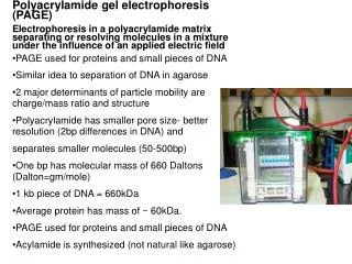
Polyacrylamide Gel Electrophoresis. By Angel Luis Vázquez Maldonado November 8 th , 2018. Gel Electrophoresis and its Purpose. Electrophoresis is derived from Greek Electro – refers to the electrical current that adds energy to the electrons of the molecule’s atoms

E N D
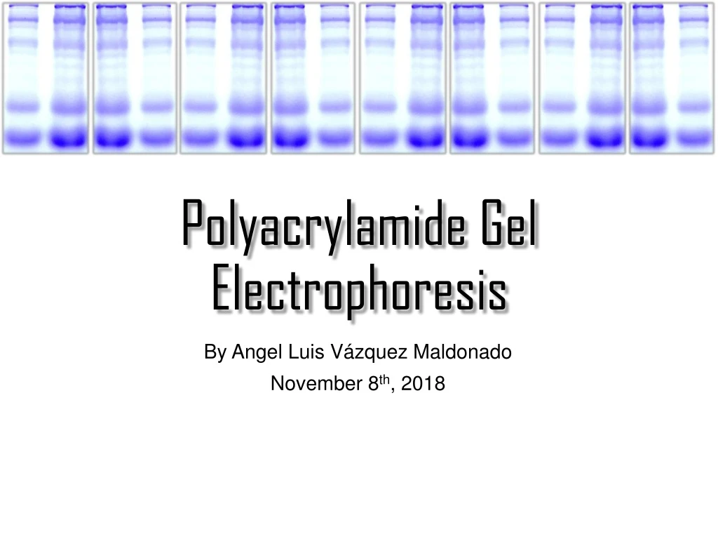
Polyacrylamide Gel Electrophoresis By Angel Luis Vázquez Maldonado November 8th, 2018
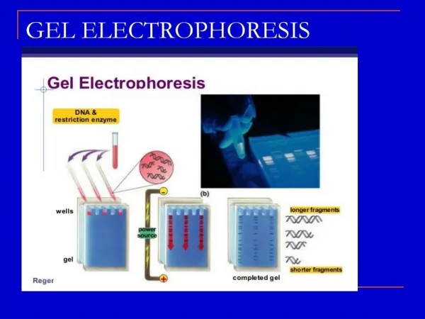
Gel Electrophoresis and its Purpose • Electrophoresis is derived from Greek • Electro – refers to the electrical current that adds energy to the electrons of the molecule’s atoms • phoresis– refers to the movement of the particles. • A technique used for separating macro molecules such as proteins, RNA and DNA. • This is done using a gel matrix through which an electric current is applied. • Utilizes an electrical current to separate target molecules based on size, charge and other physical properties, through a porous gel matrix. Oliver Smithies Dephoff, J. The Purpose of Electrophoresis. https://sciencing.com/purpose-electrophoresis-5426937.html
History of Gel Electrophoresis • Arne Tiselius is the pioneer of electrophoresis thanks to his successful protein separation experiment in 1937. • First gel electrophoresis technique was developed by Oliver Smithies in the 1950s • Starch Gel Electrophoresis Arne Tiselius Oliver Smithies Smithies, O.; Coffman, T. Annual Review of Physiology2015, 77 (1), 1–11.
Fundamentals of Electrophoresis Cathode (-) Anode (+)
Types of Gel Electrophoresis • Depending on use and application, there are different medias that can be used for the electrophoresis. • Paper – small molecules and amino acids • Polyacrylamide gel – proteins and nucleic acids • Agarose gel – very large proteins and nucleic acids • Starch gel – proteins and nucleic acids • Gel electrophoresis can be divided by two categories. • One dimension • SDS-PAGE • Native-PAGE • Isoelectric Focusing (IEF) • Two dimensions • 2D-PAGE Flint, S.; Hartley, N.; Avery, S.; Hudson, J. Letters in Applied Microbiology1996, 22 (1), 16–17.
Gel Composition • Media composition for SDS-PAGE and Native PAGE gels is acrylamide, which forms cross-linked polymers of polyacrylamide. • Gel is composed by two layers: • Stacking Gel • Separating Gel • Stacking promotes better resolution and sharper bands in separating gel. Stacking Gel Separating Gel Thermofisher. Overview of Protein Electrophoresis. https://www.thermofisher.com/pr/en/home/life-science/protein-biology/overview-electrophoresis.html.
Polyacrylamide Polymer for Gels Acrylamide • Most commonly used for proteins. • Can offer small pore sizes. • Chemically inert. • Rapidly formed. N,N’-methylene bisacrylamide Sulfate radicals Persulfate ion Thermofisher. Overview of Protein Electrophoresis. https://www.thermofisher.com/pr/en/home/life-science/protein-biology/overview-electrophoresis.html.
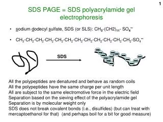
Sodium Dodecyl Sulphate Polyacrylamide Gel Electrophoresis (SDS-PAGE) • A method to separate proteins based strictly by their mass. • With the use of the strong protein-denaturing detergent SDS, the secondary and tertiary structures are disrupted by breaking hydrogen bonds and unfolding the protein. Hegyi, G. Polyacrylamide gel electrophoresis. http://elte.prompt.hu/sites/default/files/tananyagok/IntroductionToPracticalBiochemistry/ch07s03.html.
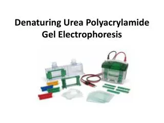
Gel Preparation for SDS-PAGE • Assemble glass plates on casting frames and place on casting stand • Make sure no leaking is happening through glass plates. • Prepare separating gel solution • Add solution to glass plates and allow to solidify. (~3-4 mL; must leave space for stacking gel) • Add ~200 uL of Ethanol 35% on top so that the gel polymerizes smoothly. • Prepare stacking gel solution • Remove excess of ethanol on the glass plates and then add the stacking gel (~ 2 mL or until glass plate is almost full). Leave space for comb! • Insert well-forming comb without trapping air bubbles. Allow to gelate before using. Bio-Rad. “A Guide to Polyacrylamide Gel Electrophoresis and Detection.” Bio-Rad Laboratories.
Separating and Stacking Recipes *Separating=Resolving **TEMED goes last Bio-Rad. “A Guide to Polyacrylamide Gel Electrophoresis and Detection.” Bio-Rad Laboratories.
Sample Preparation and Loading • Prepare a sample staining solution. • Add 10% of reducing agent 2-mercaptoethanol to staining solution (Coomassie Brilliant Blue) • Mix 2:1 Sample and staining solution. • Heat samples at 90 ºC for 1 minute. • Add samples to wells • 15-well gel – add 15 uL • 10-well gel – add 20 uL Bio-Rad. “A Guide to Polyacrylamide Gel Electrophoresis and Detection.” Bio-Rad Laboratories.
Sample Run and Staining/Destaining • After sample loading, gel is ready for run. • Running conditions: 200 V • Running time: ~40-50 min • Remove gel from glass plates and wash 3 times with distilled water. • Soak on Coomassie Brilliant Blue Stain and leave overnight gently agitating. • 0.1% Coomassie, 10% acetic acid, 40% methanol • Wash gel 3 times with distilled water. • Soak on destaining solution, add two kim wipes, microwave for 10 seconds and gently agitate for ~30–60 minutes • 10% acetic acid, 20% methanol • Analyze gel! Bio-Rad. “A Guide to Polyacrylamide Gel Electrophoresis and Detection.” Bio-Rad Laboratories.
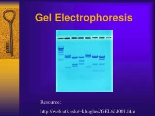
Destained SDS-PAGE Gel Example Ladder F5 F6 F9 F4 F1 F2 F7 F3 200 kDa 150 kDa 100 kDa 75 kDa 50 kDa 37 kDa 25 kDa
Native-Page • A method used to separate proteins in their native states according to difference in their charge density. • Native state means that proteins are in properly folded state, not denatured. • Mobility depends on protein’s charge and hydrodynamic size. • Proteins remains enzymatically active after separation. • Can be used as a preparation procedure for protein purification. Large, high (+) charge Large, low (+) charge Small, high (+) charge Small, low (+) charge Bio-Rad. “A Guide to Polyacrylamide Gel Electrophoresis and Detection.” Bio-Rad Laboratories.
Gel Preparation for Native-PAGE • Assemble glass plates on casting frames and place on casting stand • Make sure no leaking is happening through glass plates. • Prepare separating gel solution • Add solution to glass plates and allow to solidify. (~3-4 mL; must leave space for stacking gel) • Add ~200 uL of Ethanol 35% on top so that the gel polymerizes smoothly. • Prepare stacking gel solution • Remove excess of ethanol on the glass plates and then add the stacking gel (~ 2 mL or until glass plate is almost full). Leave space for comb! • Insert well-forming comb without trapping air bubbles. Allow to gelate before using. Bio-Rad. “A Guide to Polyacrylamide Gel Electrophoresis and Detection.” Bio-Rad Laboratories.
Separating and Stacking Recipes *Separating=Resolving **TEMED goes last Bio-Rad. “A Guide to Polyacrylamide Gel Electrophoresis and Detection.” Bio-Rad Laboratories.
Sample Preparation and Loading • Mix 2:1 Sample and sample buffer. • Sample Buffer: 25% glycerol, 62.5 mM Tris-HCl, pH 6.8, 1% Coomassie Bright Blue • Add samples to wells • 15-well gel – add 15 uL • 10-well gel – add 20 uL Bio-Rad. “A Guide to Polyacrylamide Gel Electrophoresis and Detection.” Bio-Rad Laboratories.
Sample Run and Staining/Destaining • To prevent protein denaturation by heat, system should be placed on ice. • After sample loading, gel is ready for run. • Running conditions: 100 V • Running time: ~90-120 min • Remove gel from glass plates and wash 3 times with distilled water. • Soak on Coomassie Brilliant Blue Stain and leave overnight gently agitating. • 0.1% Coomassie, 10% acetic acid, 40% methanol • Wash gel 3 times with distilled water. • Destain on water with kim wipes. • Analyze gel! Bio-Rad. “A Guide to Polyacrylamide Gel Electrophoresis and Detection.” Bio-Rad Laboratories.